International Journal of Systematic and Evolutionary Microbiology (2009), 59, 2977–2986
Janibacter hoylei sp. nov., Bacillus isronensissp. nov. and Bacillus aryabhattai sp. nov., isolatedfrom cryotubes used for collecting air from theupper atmosphere
S. Shivaji,1 Preeti Chaturvedi,1 Zareena Begum,1 Pavan Kumar Pindi,1R. Manorama,1 D. Ananth Padmanaban,2 Yogesh S. Shouche,3Shrikant Pawar,3 Parag Vaishampayan,3 C. B. S. Dutt,4 G. N. Datta,4R. K. Manchanda,5 U. R. Rao,4 P. M. Bhargava6 and J. V. Narlikar7
1Centre for Cellular and Molecular Biology, Uppal Road, Hyderabad 500 007, India
2Microbial Type Culture Collection (MTCC), Institute of Microbial Technology, Sector 39A,
3Microbial Culture Collection, National Centre for Cell Science, Pune University Campus,
4ISRO Headquarters, Department of Space, Bangalore 560 023, India
5Tata Institute of Fundamental Research, Homi Bhabha Road, Colaba, Mumbai 400 005, India
6Anveshna, 12-13-100, Lane No. 1, Street No. 3, Tarnaka, Hyderabad 500 017, India
7Inter-University Centre for Astronomy and Astrophysics, Ganeshkhind, Post Bag 4, Pune 411 007,
Three novel bacterial strains, PVAS-1T, B3W22T and B8W22T, were isolated from cryotubesused to collect air samples at altitudes of between 27 and 41 km. Based on phenotypiccharacteristics, chemotaxonomic features, DNA–DNA hybridization with the nearest phylogeneticneighbours and phylogenetic analysis based on partial 16S rRNA gene sequences (PVAS-1T,1196 nt; B3W22T, 1541 nt; B8W22T, 1533 nt), the three strains were identified as representingnovel species, and the names proposed are Janibacter hoylei sp. nov. (type strain PVAS-1T5MTCC 8307T 5DSM 21601T 5CCUG 56714T), Bacillus isronensis sp. nov. (type strainB3W22T 5MTCC 7902T 5JCM 13838T) and Bacillus aryabhattai sp. nov. (type strain B8W22T5MTCC 7755T 5JCM 13839T).
Evidence for the occurrence of micro-organisms in the
atmosphere (Bruch, 1967; Greene et al., 1964; Lysenko, 1979;
upper atmosphere at altitudes of 17–85 km (Bruch, 1967;
Rogers & Meier, 1936; Harris et al., 2002). In these earlier
Greene et al., 1964; Lysenko, 1979), obtained using a
reports, the emphasis was on detection of micro-organisms
meteorological rocket (Imshenetsky et al., 1978; Lysenko,
rather than their identification. Wainwright et al. (2003)
1979), a specially designed direct-flow sampler (Greene
reported the presence of two bacterial species (Bacillus simplex
et al., 1964; Rogers & Meier, 1936) and cryosamplers sent
and Staphylococcus pasteurii) and a fungus (Engyotontium
up on a balloon (Harris et al., 2002; Wainwright et al.,
album), whereas Shivaji et al. (2006) identified four novel
2003; Shivaji et al., 2006), has established unequivocally
species of Bacillus, Bacillus aerius, B. aerophilus, B. strato-
that life forms are present in the upper atmosphere. To
sphericus and B. altitudinis, from cryogenic tubes used to
date, only a few studies have been published on the
collect air samples at altitudes of 24, 28 and 41 km.
quantity and nature of the micro-organisms in the upper
In this paper, a polyphasic taxonomic approach was usedto characterize 12 bacterial strains isolated from cryotubes
The GenBank/EMBL/DDBJ accession numbers for the 16S rRNA gene
that were used to collect air at altitudes of between 27 and
sequences of strains PVAS-1T, B3W22T and B8W22T are DQ317608,EF114311 and EF114313.
41 km, during a balloon flight. Three of the 12 bacterialisolates, strains PVAS-1T, B3W22T and B8W22T, represent
Polar lipid profiles of the novel strains and related type strains andresults of UV-sensitivity experiments are available as supplementary
three novel species of the genera Bacillus (B3W22T and
material with the online version of this paper.
Detection of viable bacteria from air collected inthe cryotubes
The balloon with the cryosampler payload used forcollecting high-altitude air samples (altitude 20–41.4 km)
All the work related to detection of microbes in air samples
was launched on 20 April 2005 from the National Scientific
from the cryotubes was carried out in a room with very
Balloon Facility of the Tata Institute of Fundamental
little human traffic and inside a laminar flow hood
Research at Hyderabad, India. The cryogenic sampler,
available exclusively for the handling of the probes. The
comprising a 16-cryoprobe assembly, is similar to one used
probes were transferred to the laminar flow hood one at a
previously (Wainwright et al., 2003; Shivaji et al., 2006). All
time and, prior to the transfer, the surface of the cryotubes
16 cryosampling tubes were autoclaved at 120 uC for 4 h and
was cleaned and sterilized with alcohol. All instruments
integrated into the payload in a tissue-culture laboratory
used to unscrew the outlet valve of the cryotube such as
which was UV-sterilized overnight. The cryotubes were also
spanners and screwdrivers were surface-sterilized with
checked for sterility after the assembly. For this purpose,
alcohol and flame-heated prior to use. The outlet valve of
three cryotubes were disconnected from the assembly,
the cryotube was then connected to a series of two
opened in a laminar flow hood, rinsed with sterile phosphate
Millipore filtration units using sterile tubing and the air
buffer (0.1 M, pH 7.2) and plated on Luria–Bertani (LB)
was sequentially filtered through a 0.45 mm filter (Millipore
agar and incubated at 37 uC. No colonies had appeared after
cat. no. HAWP 04700) and a 0.22 mm filter (Millipore cat.
1 month, indicating that the cryotubes were sterile.
no. GSWP 04700) under aseptic conditions in the laminar
Therefore, the isolates reported in Table 1 should have
flow hood. The 0.45 mm and 0.22 mm filters were then used
originated from the air samples collected at the given
for detection of bacteria. For this purpose, each filter was
altitudes. Cryotubes 1, 3, 5, 7, 9, 12, 13 and 15 were
cut into quarters and one of the quarters was transferred to
examined at the Centre for Cellular and Molecular Biology,
a nutrient agar plate (0.5 % peptone, 0.3 % beef extract,
Hyderabad, while cryotubes 2, 4, 6, 8, 10 and 14 were
0.5 % NaCl and 1.5 % agar, w/v) and incubated at 15 uC.
similarly studied at the National Centre for Cell Sciences,
After about 10 days, if no growth was observed, the filter
Pune. Care was taken that the two laboratories followed
was transferred to an LB agar plate (1 % tryptone, 0.5 %
similar protocols, and there was frequent interaction and
yeast extract, 1.0 % NaCl and 2.0 % agar, w/v; pH 7.2) or
discussion between the two groups to ensure homogeneity
an oligotrophic LB agar plate (1/10 LB) and incubated at
15 uC for a further 15 days. Subsequently, if no colonies
Table 1. Bacteria isolated from cryotubes used for collecting air at altitudes of 20–41.4 km
With the exception of strain PVAS-1T, which was isolated from an air sample, all the strains were isolated from washes of the cryotubes.
International Journal of Systematic and Evolutionary Microbiology 59
Novel Janibacter and Bacillus species from the stratosphere
appeared, the filters were incubated on LB agar at 25 uC for
minimal salts agar, blood agar and Sabouraud agar, and
up to a month. It should be noted that, when hydrophobic
incubated under the conditions described above. The plates
0.45 mm (Millipore cat. no. HVHP 04700) and 0.22 mm
were observed for the appearance of colonies over a period
(cat. no. GVHP 04700) filters were used for filtering the air,
a thin layer of medium of the same composition was
From the 15 cryotube wash samples, 12 bacterial colonies
applied to the surface of the plates before the filters were
were detected (Table 1). Nine (PVAS-2, -3, -4, -5, -6, -8
placed on the surface of the medium.
and -10 and B5W22-1 and B5W22-2) showed more than
Two of the remaining quarters of the filter were transferred
98 % 16S rRNA gene sequence similarity to reported
to minimal salts agar. This medium contained (per 100 ml)
species (Table 1). Therefore, further attempts were not
20 ml of a 3.39 % (w/v) sodium dihydrogen phosphate
made to characterize them to the species level.
solution, 20 ml 1.5 % (w/v) potassium phosphate, 20 ml0.25 % (w/v) NaCl, 20 ml 0.5 % (w/v) ammonium chloride
and 18 ml Millipore water. The final concentration of agar
was 2 %. The medium was also supplemented with 2 mlsterile 20 % glucose and 0.2 ml sterile 0.1 M MgSO4
Morphological, growth and biochemical studies of viable
solution; the pH was adjusted to 7. This medium was
colonies were performed using standard methods (Holding
used for the cultivation of organisms which require low
& Collee, 1971; Smibert & Krieg, 1994). LB agar was used
nutrients. Blood agar [2.3 % (w/v) peptone, 0.1 % (w/v)
for growth and maintenance and for the determination of
corn starch, 0.5 % (w/v) NaCl and 1.5 % (w/v) agar, to
phenotypic and chemotaxonomic characteristics of all
which 5 % (v/v) defibrinated blood was added (after
strains except PVAS-2 and -3. Strains PVAS-2 and -3 were
autoclaving and cooling the medium)] was used for the
grown on a medium containing 0.25 % (w/v) K2HPO4,
cultivation of organisms which require rich media. Plates
0.225 % (w/v) NaH2PO4, 0.05 % (w/v) (NH4)2SO4, 0.02 %
of minimal salt agar and blood agar were incubated at
(w/v) MgSO4 . 7H2O and 0.1 % (v/v) methanol. The shape,
30 uC. One quarter of the filter was also incubated at 30 uC
size and motility of the strains were ascertained using a
on Sabouraud agar (1.0 % enzymic digest of casein, 4 %
Leitz Diaplan phase-contrast microscope with an oil-
glucose and 1.5 % agar, w/v) to facilitate fungal growth
immersion objective (6100). The sensitivity of the cultures
to antibiotics was determined by using antibiotic discs(HiMedia). Utilization of various carbon compounds as
Of the 15 cryotubes from which air was sampled, only a
sole carbon sources was tested in mineral liquid medium
single novel bacterial colony was detected, in cryotube 11,
supplemented with 0.2 % filter-sterilized carbon source as
corresponding to air collected at an altitude of 40–41.4 km
described previously (Shivaji et al., 2006). Fatty acid and
(Table 1). When placed on minimal salts agar, the filter
lipid composition (Sato & Murata, 1988; Kiran et al.,
paper from cryotube 11 showed a single cream-coloured
2004), DNA G+C content (Shivaji et al., 1992, 2005) and
colony after 24 days of incubation at 30 uC, and it was
DNA–DNA hybridization (Tourova & Antonov, 1987;
Shivaji et al., 1992) were determined according to standard
During the filtration process, plates containing fungal and
procedures. Analyses of polar lipids, respiratory quinones
bacterial media were left open in the laminar flow hood
and meso-diaminopimelic for PVAS-1T were carried out by
throughout the operation to check the sterility of the hood.
the Identification Service of the DSMZ (Braunschweig,
These plates were incubated at 25 uC (for fungi) or 37 uC
Germany). Isoprenoid quinones were also extracted
(bacteria) for 7 days prior to use, to confirm the sterility of
according to the method of Collins et al. (1977), separated
the plates. Colonies were not detected on any of these
by HPLC and identified as described previously (Reddy
plates, indicating that the sampling was done under sterile
et al., 2003). Peptidoglycan was prepared and analysed
according to the method described by Komagata & Suzuki(1987).
Detection of viable bacteria from washes of
Detailed phenotypic and chemotaxonomic characteristics
(listed in Tables 2, 3 and 4 and in the species descriptions)were compared with those of Bacillus silvestris DSM
It seemed possible that some bacteria might have remained
12223T, Bacillus megaterium MTCC 428T, Janibacter
at the bottom of the tubes or attached to their polished
anophelis CCUG 49715T, Janibacter terrae DSM 13876T,
walls and thus escaped detection. To check this possibility,
Janibacter melonis DSM 16063T, Janibacter limosus DSM
all 15 empty cryotubes were injected with 200 ml sterile
11140T and Janibacter corallicola DSM 18906T.
phosphate buffer (0.1 M, pH 7.2) and agitated at 22 uC for6 h in a shaker. The liquid was then removed using sterile
Cells of strain PVAS-1T are Gram-positive, coccoid, non-
tubing and a syringe and filtered through a 0.22 mm
endospore-forming and non-motile and occur singly or in
Millipore filter (47 mm diameter). These filters were each
clumps. Some biochemical characteristics are listed in
cut aseptically into four sectors and each sector was placed
Table 2. meso-Diaminopimelic acid is the diagnostic
on a plate containing one of four different media, LB agar,
diamino acid in the cell-wall peptidoglycan and iso-C16 : 0
Table 2. Physiological properties that differentiate strain
C16 : 1D9c as the predominant fatty acids (Tables 3 and 4).
PVAS-1T and the type strains of other species of the genus
The polar lipid profile of strain B3W22T contains
(PE), phosphatidylserine (PS) and one unidentified lipid,
Strains: 1, Janibacter hoylei sp. nov. PVAS-1T (data from this study);
whereas that of strain B8W22T has only PG and PE. MK-6,
2, Janibacter anophelis DSM 18333T; 3, Janibacter terrae DSM 13876T;
MK-7 and MK-8 are the menaquinones in strain B3W22T
4, Janibacter limosus DSM 11140T (unless indicated, data in columns
whereas, in B8W22T, only MK-7 was present (Table 3).
2–4 from Ka¨mpfer et al., 2006); 5, Janibacter melonis DSM 16063T
The DNA G+C contents of the two strains are 38–
(Yoon et al., 2004); 6, Janibacter corallicola DSM 18906T (Kageyama et
40 mol%. These characteristics indicated that strains
al., 2007). All strains were negative for growth at 40 uC, utilization of
B3W22T and B8W22T are members of the genus Bacillus
N-acetylglutamic acid, haemolysis, motility, methyl red test and H2S
production and positive for catalase, starch hydrolysis and gelatinhydrolysis. All strains tolerate up to 6 % NaCl. +, Positive; 2,negative; W, weakly positive; d, delayed reaction; TSBA, tryptic soy
broth agar; CASO agar, casein-peptone-soymeal peptone agar.
The 16S rRNA gene was amplified from genomic DNA,purified and sequenced as described previously (Shivaji et
al., 2000; Pidiyar et al., 2004). To ascertain the phylogenetic
affiliation of the novel strains, the almost-complete 16S
rRNA gene sequences of the bacterial isolates were manually
corrected and aligned using CLUSTAL_X (Thompson et al.,
1994). Phylogenetic trees were constructed based on
neighbour-joining (Saitou & Nei, 1987) and maximum-
parsimony methods using MEGA 3.1 (Kumar et al., 2004).
Evolutionary distances were determined with Kimura’s two-
parameter model (Kimura, 1980). Bootstrap analysis
(Felsenstein, 1993) was performed for 1000 replications.
Reference sequences were retrieved from GenBank under the
accession numbers indicated on the trees.
Phylogenetic analysis of the 16S rRNA gene sequence of
strain PVAS-1T (1196 bp) indicated that its closest relatives
were Janibacter anophelis CCUG 49715T (98 % similarity)
and Janibacter terrae DSM 13953T (98 %) (Lang et al.,
2003). Both the neighbour-joining tree (Fig. 1) and the
maximum-parsimony tree (not shown) revealed that strain
PVAS-1T clustered most closely with these strains. DNA–
DNA relatedness of PVAS-1T with Janibacter terrae DSM
13953T was 29.8 % and with Janibacter anophelis DSM
18333T was 13.7 %, and the reciprocal reactions in both
cases gave values of about 9–10 % . Thus, strain PVAS-1T
represents a novel species of the genus Janibacter, for whichthe name Janibacter hoylei sp. nov. is proposed.
*Data generated for this study by the Identification Services of the
Strains B3W22T and B8W22T are related at the 16S rRNA
gene sequence level (91 % similarity), but the sequencesdiffer by more than 2.5 %, indicating that they probablyrepresent different species. BLASTN analysis indicated that
(50.4 %), iso-C18 : 0 (5.1 %), 10-methyl C17 : 0 (10.9 %),
the nearest phylogenetic neighbour of B3W22T is Bacillus
C17 : 1D9c (12.1 %) and C18 : 1D9c (6.0 %) are the predom-
silvestris H3-23T (99.5 % similarity), with which it forms a
inant fatty acids. Mycolic acids are absent. The predom-
clade in the phylogenetic tree, and it is separated from
inant isoprenoid quinone present is MK-8(H4). The DNA
Caryophanon latum DSM 14151T and Caryophanon tenue
G+C content is 72.8 mol%. These characteristics and
DSM 14152T, with which it exhibits about 97 and 96 %
phylogenetic analysis of the 16S rRNA gene sequence
similarity, respectively (Fig. 2). The members of the genus
indicated that PVAS-1T is a member of the genus
Caryophanon are characterized by the presence of slightly
curved to straight trichomes, and are thus clearly differentfrom B3W22T.
Cells of strains B3W22T and B8W22T are Gram-positive,rod-shaped, endospore-forming and catalase-positive, with
DNA–DNA hybridization studies indicated that the
C15 : 0, C16 : 0, iso-C15 : 0, anteiso-C14 : 0, anteiso-C16 : 0 and
relatedness between B3W22T and Bacillus silvestris DSM
International Journal of Systematic and Evolutionary Microbiology 59
Novel Janibacter and Bacillus species from the stratosphere
gravitophotophoresis (Rohatschek, 1996). Finally, in 1982,
Furthermore, these two strains exhibit phenotypic and
Hoyle and Wickramasinghe proposed the theory of
chemotaxonomic differences (Tables 3 and 4) that imply
‘Panspermia’ (Hoyle & Wickramasinghe, 1986, 1993,
that B3W22T represents a novel species; the name proposed
Description of Janibacter hoylei sp. nov.
In the phylogenetic tree, strain B8W22T forms a robustclade with its nearest phylogenetic neighbour Bacillus
Janibacter hoylei (hoy9le.i. N.L. gen. n. hoylei of Hoyle,
megaterium IAM 13418T (Fig. 2), with which it shows
named after Sir Fred Hoyle, the famous English astro-
99.7 % similarity at the 16S rRNA gene sequence level.
Despite this high similarity, it is observed that, at the whole
Colonies on LB agar are creamish, entire, round and 1–
genome level, the DNA–DNA relatedness between B8W22and Bacillus megaterium MTCC 428T is only 35 %
2 mm in diameter. Cells stain Gram-positive and show
(reciprocal reaction 43 %), indicating that B8W22T could
oxidative metabolism; they are non-motile, non-endo-
be assigned to a novel species. Furthermore, B8W22T and
spore-forming cocci, 0.4–0.7 mm in diameter, that occur
Bacillus megaterium MTCC 428T differ in their phenotypic
singly or in clumps. Good growth (visible colonies with a
and chemotaxonomic characteristics (Tables 3 and 4),
diameter of 1 mm) occurs after 2 days of incubation on
implying that B8W22T does indeed represent a novel
nutrient agar at 25–30 uC. Grows in LB broth at 20–40 uC
species, for which the name proposed is Bacillus aryabhat-
and at pH 5–10, with optimum growth at 30 uC and pH 9.
tai sp. nov. Similarity of B8W22T to other Bacillus species
Grows in the presence of 5 % (w/v) NaCl in LB broth and
at the 16S rRNA gene sequence level was less than 95 %.
exhibits weak growth in LB broth containing 10 % NaCl. Resistant to UV radiation. Catalase- and oxidase-positive;H2S is not produced. Voges–Proskauer negative, indole-
negative and reduces nitrate to nitrite. Results of carbon
source utilization tests are shown in Table 2. Does not
sensitivity to UV radiation was determined by exposure to
produce acid from alcohols or the following sugars: D-
a UV lamp (UV-B, 15 W64; Sankyo Denki) as described
glucose, D-fructose, D-galactose, lactose, maltose, cello-
previously (Shivaji et al., 2006). Cultures of Bacillus
biose, D-mannitol, D-mannose, raffinose, L-rhamnose,
Micrococcus luteus MTCC 106T, Janibacter limosus DSM
Diaminopimelic acid is the diagnostic diamino acid in
11140T and Janibacter terrae DSM 13876T were used as
the cell-wall peptidoglycan. The main fatty acids present
controls in these experiments. The results indicate that
are iso-C16 : 0, 10-methyl C17 : 0, C18 : 1D9c and C17 : 1D9c. The
strains B3W22T, B8W22T and PVAS-1T are more resistant
predominant isoprenoid quinone present is MK-8(H4). In
to UV irradiation than their nearest phylogenetic neigh-
whole-cell hydrolysates, ribose and glucose are present, but
bours (Supplementary Table S1, available in IJSEM
arabinose, galactose and mannose are absent. Mycolic acids
are absent. The DNA base composition of the type strain is72.8 mol% G+C.
The type strain is PVAS-1T (5MTCC 8307T 5DSM21601T 5CCUG 56714T), isolated from an air sample
The bacterial strains identified are unlikely to be laboratory
collected at an altitude of 40–41.4 km using a cryosampler.
contaminants, because no such cultures were handled inthe laboratory. The control plates that were exposed to theair flow of the laminar flow hood during the entire air
Description of Bacillus isronensis sp. nov.
Bacillus isronensis (is.ro.nen9sis. N.L. masc. adj. isronensis
Furthermore, all possible precautions were taken to rule
arbitrary name pertaining to ISRO, the acronym of the
out contamination of the cryotubes throughout the
Indian Space Research Organization, which largely funded
assembly and deployment stages of the experiment; it is
the studies in which the type strain was isolated).
unlikely that they came from the cryotube assembly facilityand survived the transit through the stratosphere, because
Colonies on nutrient agar are white, entire, round and 3–
the control cryotubes were completely sterile. It has been
4 mm in diameter. Cells produce round terminal
argued that micro-organisms can travel across interplan-
endospores and are motile. Grows at 5–37 uC and
etary space during routine meteoritic exchanges between
pH 6–10. Does not grow at 42 uC or at pH 4 or 11.
the Earth and Mars (Gladman et al., 1996; Mileikowsky
Tolerates up to 5.8 % NaCl. Resistant to UV radiation.
et al. 2000; Nicholson et al., 2000). Furthermore, it is
Grows on peptone. Positive for oxidase, lipase, gelatinase,
possible that micro-organisms reach the stratosphere from
starch hydrolysis, caseinase, tryptophan deamination,
the Earth as a result of volcanic eruptions, the updraft
nitrate reduction and indole production. Produces acid
caused by blue lightning strikes (Pasko et al., 2002),
from cellobiose and utilizes a number of sugars, amino
thunderstorms and forest fires (Fromm et al., 2004) and
acids and other carbon compounds as sole carbon sources
Table 3. Physiological properties that differentiate strains B3W22T and B8W22T and their nearest phylogenetic neighbours
Strains: 1, Bacillus isronensis sp. nov. B3W22T; 2, Bacillus silvestris DSM 12223T; 3, Bacillus aryabhattai sp. nov. B8W22T; 4, Bacillus megateriumMTCC 428T. All four strains have Gram-positive, motile, rod-shaped cells that produce endospores, grow at 20–37 uC and pH 6–10, tolerate 5.8 %NaCl and grow in peptone broth and are positive for catalase and b-galactosidase but do not grow at pH 4 or 11 and are negative for citrateutilization, the methyl red test and H2S production. None of the strains produces acid from sucrose or inulin or uses dextran, citric acid, cellulose,glycogen, thioglycolate, hydroxybutyric acid, salicin, D-glucuronic acid, L-aspartic acid, sodium succinate, valeric acid, L-cysteine, L-tyrosine, L-phenylalanine, L-proline or L-histidine as sole carbon sources. All four strains utilize glycerol, myo-inositol, methyl a-D-galactoside, L-ornithine and
L-creatinine as sole carbon sources, are resistant to discs containing colistin (10 mg) and are sensitive to discs containing norflaxacin (10 mg),
tobramycin (15 mg), lomefloxacin (30 mg), amikacin (30 mg), roxithromycin (15 mg), ciprofloxacin (30 mg), nitrofurantoin (300 mg), cefoperazone(75 mg), vancomycin (30 mg), lincomycin (15 mg), cephotaxime (30 mg), kanamycin (30 mg), novobiocin (30 mg), chloramphenicol (30 mg),ampicillin (25 mg), tetracycline (30 mg), bacitracin (10 mg), gentamicin G (30 mg), polymyxin B (50 mg), oleandomycin (15 mg), spectinomycin(100 mg), rifampicin (30 mg), erythromycin (15 mg) and carbenicillin (100 mg). +, Positive; 2, negative; V, variable; W, weak; S, sensitive;R, resistant.
Trehalose, maltose, adonitol, erythritol,
International Journal of Systematic and Evolutionary Microbiology 59
Novel Janibacter and Bacillus species from the stratosphere
L-arabinose, melibiose, sucrose, D-mannitol,raffinose, D-mannose, D-xylose,
L-Sorbose, D-fructose, trehalose, L-xylose,
lactose, maltose, propionate, starch, methyla-D-mannoside, polyethylene glycol,sodium acetate, potassium acetate,amygdalin, xylitol, erythritol
Lactic acid, adonitol, N-acetylglucosamine,
L-Serine, L-threonine, L-arginine, L-glutamic
Cefazolin (30 mg), co-trimoxazole (25 mg)
Streptomycin (25 mg), nalidixic acid (30 mg)
*DAP, Diaminopimelic acid. DDPG, Diphosphatidylglycerol; PE, phosphatidylethanolamine; PG, phosphatidylglycerol; PS, phosphatidylserine; UL, unknown lipid.
(Table 3). The type strain is sensitive to a number of
The type strain is B3W22T (5MTCC 7902T 5JCM
antibiotics (Table 3). The fatty acids present are C15 : 0,
13838T), isolated from a cryogenic tube used for collecting
C16 : 0, C17 : 0, iso-C13 : 0, iso-C15 : 0, anteiso-C14 : 0, anteiso-
an air sample from an altitude of 27–30 km.
C16 : 0, C16 : 1D9c, C16 : 1D11 and C18 : 1D9 (Table 4). Theinterpeptide bridge of the cell-wall peptidoglycan consists
Description of Bacillus aryabhattai sp. nov.
of Lys–Glu–Ala. The lipids present are PG, PE, PS and anunidentified lipid. MK-6, MK-7 and MK-8 are the
Bacillus aryabhattai (a.ry.a.bhat9ta.i. N.L. gen. n. aryabhat-
menaquinones present. The DNA G+C content of the
tai of Aryabhatta, named after the renowned Indian
Table 4. Total fatty acid compositions of strains B3W22T andB8W22T and related type strains
Strains: 1, Bacillus isronensis sp. nov. B3W22T; 2, Bacillus silvestrisDSM 12223T; 3, Bacillus aryabhattai sp. nov. B8W22T; 4, Bacillusmegaterium MTCC 428T. Data are percentages of total fatty acids andwere obtained in this study; strains were grown on nutrient agar (1 %beef extract, 1 % peptone, 0.5 % NaCl and 1.5 % agar; pH 7.2) andincubated overnight at 30 uC.
Colonies on nutrient agar are peach-coloured, entire,round, flat and 5–8 mm in diameter. Cells produce
Fig. 2. Neighbour-joining phylogenetic tree reconstructed on the
ellipsoidal and central endospores and are motile.
basis of 16S rRNA gene sequences showing the phylogenetic
Growth occurs at 10–37 uC and pH 6–10. Does not grow
relationships between strains B3W22T and B8W22T and closely
at 42 uC or at pH 4 or 11. Tolerates up to 11.6 % NaCl.
related species of the genera Bacillus, Caryophanon and
Resistant to UV radiation. Grows on peptone. Positive for
Lysinibacillus. The sequence of Microbacterium indicum DSM
phosphatase, DNase, tryptophan deamination, ornithine
20030T (GenBank accession no. AJ536198; not shown) was
decarboxylase and malonate utilization (Table 3). Produces
used as an outgroup. Bootstrap values (expressed as percentages
acid from various carbon sources and utilizes a number of
of 1000 replications) .50 % are given at nodes. Bar, 2
sugars, amino acids and other carbon compounds as sole
substitutions per 100 nucleotide positions.
carbon sources (Table 3). The type strain is sensitive to anumber of antibiotics (Table 3). The fatty acids present areC14 : 0, C15 : 0, C18 : 0, iso-C15 : 0, anteiso-C14 : 0, anteiso-C16 : 0and C16 : 1D9c (Table 4). The interpeptide bridge of the cell-wall peptidoglycan consists of meso-diaminopimelic acid–Ala–Glu. The lipids present are PG and PE. MK-7 is themenaquinone present. The DNA G+C content of the typestrain is 38 mol%.
The type strain is B8W22T (5MTCC 7755T5JCM 13839T),
Fig. 1. Neighbour-joining phylogenetic tree reconstructed on the
isolated from a cryogenic tube used for collecting an air
basis of 16S rRNA gene sequences showing the phylogenetic
sample from an altitude of 40–41.4 km.
relationships between strain PVAS-1T and closely related speciesof the genus Janibacter. The sequence of Brevibacterium linensDSM 20425T (GenBank accession no. X77451; not shown) was
used as an outgroup. Bootstrap values (expressed as percentages
The present study is part of a long-term study partly conceptualized
of 1000 replications) .50 % are given at nodes. Bar, 1
and largely funded by the Indian Space Research Organization,
substitution per 100 nucleotide positions. The asterisk (*) indicates
Department of Space, Government of India. The payload envir-
that this was the only branch point that was conserved in the
onmental testing and some critical parameter evaluations were carried
out at various ISRO Centres (VSSC, LPSC and ISAC). The periodic
International Journal of Systematic and Evolutionary Microbiology 59
Novel Janibacter and Bacillus species from the stratosphere
guidance of Dr Vedachalam, Dr Lalji Singh of CCMB, Dr T. K. Alex,
Kiran, M. D., Prakash, J. S. S., Annapoorni, S., Dube, S., Kusano, T.,
LEOS, Mr V. Sundararamaiah and Mr Gnana Gandhi is acknowl-
Okuyama, H., Murata, N. & Shivaji, S. (2004). Psychrophilic
edged. We are grateful to Dr G. Madhavan Nair, Chairman, ISRO/
Pseudomonas syringae requires trans-monounsaturated fatty acid for
Secretary, DOS, for his encouragement and full support throughout
growth at higher temperature. Extremophiles 8, 401–410.
the payload development and analysis. Our special thanks go to the
Komagata, K. & Suzuki, K. (1987). Lipid and cell-wall analysis in
India Air Force for the helicopter recovery of the payload. We thank
bacterial systematics. Methods Microbiol 19, 161–207.
Dr Jean P. Euze´by, Ecole Nationale Ve´te´rinaire, Toulouse, France, forhelp with the nomenclature of the novel species. Y. S. S. would also
Kumar, S., Tamura, K. & Nei, M. (2004). MEGA3: integrated software
like to thank the Department of Biotechnology, Government of India,
for molecular evolutionary genetics analysis and sequence alignment.
Lang, E., Kroppenstedt, R. M., Swiderski, J., Schumann, P.,Ludwig, W., Schmid, A. & Weiss, N. (2003). Emended description
of Janibacter terrae including ten dibenzofuran-degrading strains andJanibacter brevis as its later heterotypic synonym. Int J Syst EvolMicrobiol 53, 1999–2005.
Bruch, C. W. (1967). Microbes in the upper atmosphere and beyond. In Airborne Microbes (Society for General Microbiology Symposium
Lysenko, S. V. (1979). Microorganisms of the upper layers of the
no. 17), pp. 345–373. Edited by P. H. Gregory & J. L. Monteith.
atmosphere. Mikrobiologiia 48, 1066–1074 (in Russian).
Cambridge: Cambridge University Press.
Mileikowsky, C., Cucinotta, F. A., Wilson, J. W., Gladman, B.,
Collins, M. D., Pirouz, T., Goodfellow, M. & Minnikin, D. E. (1977).
Horneck, G., Lindegren, L., Melosh, J., Rickman, H., Valtonen, M. &
Distribution of menaquinones in actinomycetes and corynebacteria.
Zheng, J. Q. (2000). Natural transfer of viable microbes in space.
Felsenstein, J. (1993). PHYLIP (phylogeny inference package), version
Nicholson, W. L., Munakata, N., Horneck, G., Melosh, H. J. & Setlow, P.
3.5c. Distributed by the author. Department of Genome Sciences,
(2000). Resistance of Bacillus endospores to extreme terrestrial and
University of Washington, Seattle, USA.
extraterrestrial environments. Microbiol Mol Biol Rev 64, 548–572.
Fromm, M., Bevilacqua, R., Stocks, B. & Servanckx, R. (2004). New
Pasko, V. P., Stanley, M. A., Mathews, J. D., Inan, U. S. & Wood, T. G.
directions: eruptive transport to the stratosphere: add fire-convection
(2002). Electrical discharge from thundercloud tops to the lower
to volcanoes. Atmos Environ 38, 163–165.
Gladman, B. J., Burns, J. A., Duncan, M., Lee, P. & Levinson, H. F.
Pidiyar, V. J., Jangid, K., Patole, M. S. & Shouche, Y. S. (2004).
(1996). The exchange of impact ejecta between terrestrial planets.
Studies on cultured and uncultured microbiota of wild Culex
quinquefasciatus mosquito midgut based on 16S ribosomal RNA
Greene, V. W., Pederson, P. D., Lundgren, D. A. & Hagberg, C. A.
gene analysis. Am J Trop Med Hyg 70, 597–603.
(1964). Microbiological exploration of stratosphere: results of six
Reddy, G. S. N., Prakash, J. S. S., Prabahar, V., Matsumoto, G. I.,
experimental flights. In Proceedings of the Atmospheric Biology
Stackebrandt, E. & Shivaji, S. (2003). Kocuria polaris sp. nov., an
Conference, University of Minnesota, 13–15 April 1964, pp.
199–211. Washington, DC: National Aeronautics and Space
Antarctic cyanobacterial mat sample. Int J Syst Evol Microbiol 53,
Harris, M. J., Wickramasinghe, N. C., Lloyd, D., Narlikar, J. V.,
Rogers, L. A. & Meier, F. C. (1936). In US Army Air Corps Stratosphere
Rajaratnam, P., Turner, M. P., Al-Mufti, S., Wallis, M. K., Ramadurai, S.
Flight of 1935 in the Balloon ‘‘Explorer II’’, pp. 146–151. Edited by L. J.
& Hoyle, F. (2002). Detection of living cells in stratospheric samples. In
Briggs. Washington, DC: National Geographic Society.
Instruments, Methods, and Missions for Astrobiology IV (Proceedings of
Rohatschek, H. (1996). Levitation of stratospheric and mesophilic
the SPIE Conference 2001, vol. 4495), pp. 192–198. Edited by R. B.
aerosols by gravito-photophoresis. J Aerosol Sci 27, 467–475.
Hoover, G. V. Levin, R. R. Paepe & A. Y. Rozanov. Bellingham,WA: SPIE.
Saitou, N. & Nei, M. (1987). The neighbor-joining method: a newmethod for reconstructing phylogenetic trees. Mol Biol Evol 4, 406–425.
Holding, A. J. & Collee, J. G. (1971). Routine biochemical tests. Methods Microbiol 6A, 2–32.
Sato, N. S. & Murata, N. (1988). Membrane lipids. Methods Enzymol167, 251–259.
Hoyle, F. & Wickramasinghe, N. C. (1986). The case for life as acosmic phenomenon. Nature 322, 509–511.
Shivaji, S., Ray, M. K., Rao, N. S., Saisree, L., Jagannadham, M. V.,Kumar, G. S., Reddy, G. S. N. & Bhargava, P. M. (1992).
Hoyle, F. & Wickramasinghe, N. C. (1993). Our Place in the Cosmos.
Sphingobacterium antarcticus sp. nov., a psychrotrophic bacterium
from the soils of Schirmacher Oasis, Antarctica. Int J Syst Bacteriol 42,
Hoyle, F. & Wickramasinghe, N. C. (1999). Astronomical Origins of
Life: Steps Towards Panspermia. Dordrecht: Kluwer.
Shivaji, S., Bhanu, N. V. & Aggarwal, R. K. (2000). Identification of
Imshenetsky, A. A., Lysenko, S. V. & Kazakov, G. A. (1978). Upper
Yersinia pestis as the causative organism of plague in India as
boundary of the biosphere. Appl Environ Microbiol 35, 1–5.
determined by 16S rDNA sequencing and RAPD-based genomic
Kageyama, A., Takahashi, Y., Yasumoto-Hirose, M., Kasai, H.,
fingerprinting. FEMS Microbiol Lett 189, 247–252.
Shizuri, Y. & Omura, S. (2007). Janibacter corallicola sp. nov., isolated
Shivaji, S., Chaturvedi, P., Reddy, G. S. N. & Suresh, K. (2005).
from coral in Palau. J Gen Appl Microbiol 53, 185–189.
Pedobacter himalayensis sp. nov., from the Hamta glacier located in
Ka¨mpfer, P., Terenius, O., Lindh, J. M. & Faye, I. (2006). Janibacter
the Himalayan mountain ranges of India. Int J Syst Evol Microbiol 55,
anophelis sp. nov., isolated from the midgut of Anopheles arabiensis.
Int J Syst Evol Microbiol 56, 389–392.
Shivaji, S., Chaturvedi, P., Suresh, K., Reddy, G. S. N., Rajaratnam, P.,
Kimura, M. (1980). A simple method for estimating evolutionary rates
Wainwright, M., Narlikar, J. V. & Bhargava, P. M. (2006). Bacillus aerius
of base substitutions through comparative studies of nucleotide
sp. nov., Bacillus aerophilus sp. nov., Bacillus stratosphericus sp. nov.
sequences. J Mol Evol 16, 111–120.
and Bacillus altitudinis sp. nov., isolated from cryogenic tubes used for
collecting air samples from high altitudes. Int J Syst Evol Microbiol 56,
Tourova, T. P. & Antonov, A. S. (1987). Identification of microorgan-
isms by rapid DNA-DNA hybridization. Methods Microbiol 19, 333–
Smibert, R. M. & Krieg, N. R. (1994). Phenotypic characterization. In
Methods for General and Molecular Bacteriology, pp. 607–654. Edited
by P. Gerhardt, R. G. E. Murray, W. A. Wood & N. R. Krieg.
Rajaratnam, P. (2003). Microorganisms cultured from strato-
Washington, DC: American Society for Microbiology.
spheric air samples obtained at 41 km. FEMS Microbiol Lett 218,
Thompson, J. D., Higgins, D. G. & Gibson, T. J. (1994).
improving the sensitivity of progressive multiple sequence alignment
Yoon, J. H., Lee, H. B., Yeo, S. H. & Choi, J. E. (2004). Janibacter
through sequence weighting, position-specific gap penalties and
melonis sp. nov., isolated from abnormally spoiled oriental melon in
weight matrix choice. Nucleic Acids Res 22, 4673–4680.
Korea. Int J Syst Evol Microbiol 54, 1975–1980.
International Journal of Systematic and Evolutionary Microbiology 59
November 2008 Essential oil of the Month: Vetiver Kathy Padecky, Aromatherapist, HHP Botanical Name: Vetiveria zizanoides is known as the "oil of tranquility". It is known by other names such as Vetivert, Khus-Khus and moth root. Why I love vetiver : I love the smell of vetiver for reasons I have challenges articulating. Words fail to express the depth and essence of my f
Medical Disorders and Pharmacology Instructor of Record: Lori Flynn COURSE DESCRIPTION: This course introduces the learner to important concepts related to human diseases. The most common diseases and disorders of each body system are presented along with a review of the anatomy and physiology pertinent to that system. Additionally, causes, diagnoses, treatments, and medications o
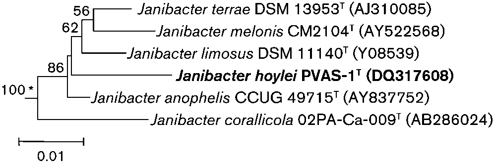
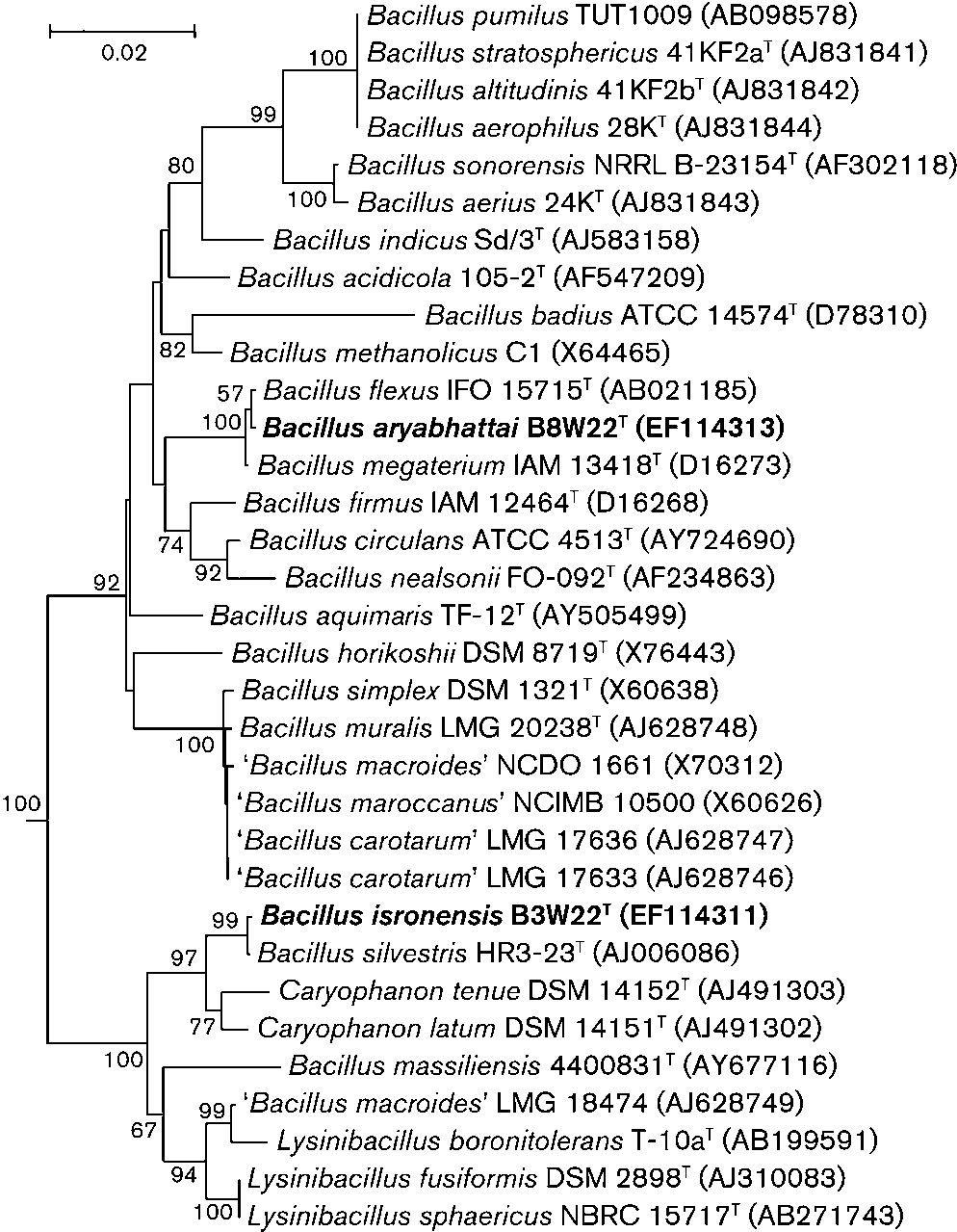 Table 4. Total fatty acid compositions of strains B3W22T andB8W22T and related type strains
Strains: 1, Bacillus isronensis sp. nov. B3W22T; 2, Bacillus silvestrisDSM 12223T; 3, Bacillus aryabhattai sp. nov. B8W22T; 4, Bacillusmegaterium MTCC 428T. Data are percentages of total fatty acids andwere obtained in this study; strains were grown on nutrient agar (1 %beef extract, 1 % peptone, 0.5 % NaCl and 1.5 % agar; pH 7.2) andincubated overnight at 30 uC.
Table 4. Total fatty acid compositions of strains B3W22T andB8W22T and related type strains
Strains: 1, Bacillus isronensis sp. nov. B3W22T; 2, Bacillus silvestrisDSM 12223T; 3, Bacillus aryabhattai sp. nov. B8W22T; 4, Bacillusmegaterium MTCC 428T. Data are percentages of total fatty acids andwere obtained in this study; strains were grown on nutrient agar (1 %beef extract, 1 % peptone, 0.5 % NaCl and 1.5 % agar; pH 7.2) andincubated overnight at 30 uC.