www.abbeyvetservices.co.uk/newsletters/oct07.htm
A Case of a Nictitans Gland Carcinoma in a Cat
• Latest news • Case of interest • Our Details
• Biopsy tips • Side Story • Journal Articles
A seven-year-old domestic short haired cat presented with a protruding mass from the third • Site Downloads
eyelid of the left orbit but also involving the upper and lower eyelid. The external aspect of the mass was approximately 18 mm in diameter and covered by hyperaemic conjunctival epithelium.
Surgical biopies of the centre of the lesion identified a subconjunctival mass extending to both JOURNAL Reviews(with e-links)
deep and lateral biopsy margins in submitted tissue sections. In one section the mass was
composed primarily of tubulo-acinar structures formed from discrete polygonal to cuboidal 1. Palmeiro BS, Morris DO, Goldschmidt MH,
cells. These cells had scant eosinophilic cytoplasm and variably sized but typically large oval Mauldin EA. Cutaneous reactive histiocytosis in
nuclei, with a single prominent nucleolus. Mitotic figures were approximately 3 per 10 hpfs. dogs: a retrospective evaluation of 32 cases. Vet
The glandular lumen occasionally contained neutrophilic or mucinous debris and neutrophils
were scattered in the scant supporting collagenous stroma.
32 cases of canine cutaneous histiocytosis were
retrospectively evaluated. Median age at onset was 4 years. Lesions included nodules and plaques affecting the head/face, trunk and limbs, and erythema, swelling and depigmentation of the nasal planum/nares. Systemic involvement was not ruled out in all cases. All dogs had complete resolution of dermatological lesions after initial treatment (median 45 days). . Median follow up was 25 months. 9 dogs had a recurrence of cutaneous histiocytosis (median days to recurrence 130 days), with 7 of 9 having more than one recurrence. At study completion, 6 dogs were deceased (no lesions at the time of death) and 26
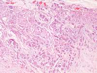
Figure 1. Neoplasm. Biopsy specimen identifies a well demarcated mass composed of
of 32 were alive with no lesions. 10 of 26 dogs
epithelial cells forming tubuloacinar structures. obj. x10. HE Stain.
were on maintenance treatment (8 tetracycline/ niacinamide, 1 azathioprine, 1 vitamin E). Previous dermatological
Following the histological diagnosis the eye was enucleated and fixed in formalin. It was detectable influence on recurrence. Recurrence was prepared for photography as shown below.
significantly more likely in dogs with nasal planum/nares lesions than dogs without these lesions. Tetracycline/niacinamide was an effective
treatment option for dogs in this study population.
2. Demko JL, Cohn LA. Chronic nasal discharge in cats: 75 cases (1993-2004). J Am Vet Med Assoc. 2007 Apr 1;230(7):1032-7. Link
This study's objective was to identify the most common aaetiologic diagnosis and any historical, physical, or other diagnostic variables associated with a definitive aetiologic diagnosis for chronic nasal discharge in cats. Design-Retrospective case
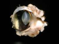
Figure 2. Globe including eyelids: The neoplasm appears to extend throughout the upper lid
series. Medical records of affected cats were
but also in the (small off-white mass) lower lid.
reviewed for information on signalment, clinical
(Gross photo courtesy of John Mould, Eye Vet Clinic, Herefordshire)
signs, duration and type of nasal discharge, results of clinical examination, laboratory findings, and advanced imaging findings. A specific aaetiologic
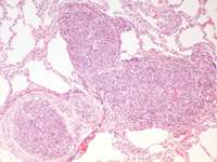
The cat was presented, four months later, inappetant and with increased respiratory effort. diagnosis for nasal discharge was identified in only There was no response to symptomatic treatment and was euthanased. Samples of liver and 36% of cats. Neoplasia (carcinoma or lymphoma) sternal lymph node were taken during a limited post mortem examination and submitted for was the most common aetiologic diagnosis. histopathology. Two sections of lung tissue displayed multifocal interstitial infiltration by Character and location of nasal discharge did not neoplastic cuboidal epithelial cells forming tubulopapillary structures. These formed discrete contribute greatly toward a specific aetiologic nodules surrounded by residual pulmonary parenchyma. Throughout the pulmonary capillaries diagnosis. Sneezing and vomiting were the most were large numbers of neoplastic cells which were also present within large muscular arteries. common concurrent clinical signs. Routine CBC, A section of sternal lymph node identified marked intrasinusoidal infiltration by similar-
serum biochemical panel, and urinalysis did not
contribute to a specific aetiologic diagnosis. An aetiologic diagnosis was more likely in older cats
and cats that underwent advanced imaging studies and nasal biopsy. Although advanced diagnostic testing, including imaging studies and biopsy, increases the likelihood of achieving an aetiologic diagnosis, the cause of chronic nasal discharge in cats often remains elusive.
3. E. M. Romansik, C. M. Reilly, P. H. Kass, P. F. Moore and C. A. London. Mitotic Index Is Predictive for Survival for Canine Cutaneous Mast Cell Tumors. Vet Pathol 44:335-341 (2007) Link
www.abbeyvetservices.co.uk/newsletters/oct07.htm
Figure 3. Lung. Tumour cells are present within the pulmonary parenchyma and within large
The purpose of the study was to evaluate the utility
of MI as a predictor of biologic behavior and survival in dogs with cutaneous mast cell tumors
Adenocarcinomas arising from the nictitans gland are a rare neoplasm in the cat. Their (MCTs). Medical records from 148 dogs with
histologically confirmed MCTs were reviewed.
behaviour is sparsely documented. They are believed to be locally invasive.
recurrence, metastatic disease, date of death/last
In dogs, where this tumour is somewhat more commonly reported, adequate excision with
follow-up, and outcome was obtained. The MI
wide margins can be curative. Whether that also applies to these tumours in the majority of correlated directly with tumor grade (P < .0001). cats is unknown. Widespread metastasis of this tumour has been reported in one case in the The median survival time for dogs with an MI ≤5 cat and obviously occured in this case. Because of the sparsity of prognostic data and because was significantly longer (70 months) than for those of the cytological malignancy of this mass, a guarded prognosis was initially given in this case with an MI >5 (2 months), regardless of grade (P and was thus justified given the outcome.
< .001). For grade II tumors with an MI ≤5, the median survival time (MST) was 70 months, compared with 5 months for those with an MI >5
(P < .001). For grade III tumors with an MI ≤5, the MST was not reached, compared with <2 months
1. D. Dubielzig (1990). Tumours of the eye. In:Meuten, D. J. (ed.). Tumors of Domestic for those with an MI >5 (P < .001). In conclusion, Animals, 4th ed. Iowa State Press, Ames, 2002., p743.
MI is a strong predictor of overall survival for dogs with cutaneous MCTs and should be included as a prognostic indicator when determining therapeutic
2. Komaromy AM, Ramsey DT, Render JA, Clark P. Primary adenocarcinoma of the gland of options.
the nictitating membrane in a cat. J Am Anim Hosp Assoc. 1997 Jul-Aug;33(4):333-6.
3. Schäffer EH, Pfleghaar S, Gordon S, Knödlseder M. Malignant nictitating membrane tumors in dogs and cats. Tierarztl Prax. 1994 Aug;22(4):382-91.
Dog flea treatments killing cats - FAB Abbey Veterinary Services website
An excerpt from "Hill's symposium on
The VPIS report highlighted the lethal risks of U.K.
permethrin based dog spot-on treatments being TQ122BG
Interstitial cystitis is apparently a misnomer. Recent research has led to
Toxic effects can also occur from cats coming into
close physical contact with dogs in the same
which it has been referred to as “feline
house (through sharing beds or grooming) that Tel: +44 (0)1626 353598
have been appropriately treated with permethrin.
These products are available in pet shops and Where we are: Multimap Link
many supermarkets, and have been mistakenly or
unwittingly used on cats, frequently causing
severe illness and even death. Cats poisoned with
permethrin may need 2-3 days of intensive
victim rather than the perpetrator of the syndrome.
The report is a review of 286 cases reported to VPIS where such canine spot-on permethrin
preparations have been used on cats. Of these
cases, 97 per cent of the cats had signs of
convulsions and 10.5 per cent of the cats died or were euthanased. Although these data are startling, the VPIS feels that they are an under-
representation of the scale of the problem.
provision of all necessary resources, control of interactions with owners, a tolerable intensity of conflict with other
The veterinary press often receives letters on the
topic from vets in practice and the Veterinary
Medicines Directorate (VMD) highlighted the problem in 2000 – ‘These spot-on products are sold through UK pet stores or supermarkets and
It is thought that a change in food also
veterinary surgeons should be alert to the
cases’ (Gray, Veterinary Record 147, p556).
suggests that cats prefer to eat individually in a quiet location where
Further info: Full FAB cats website article
they will not be startled by other animals, sudden movement, or sudden
fragmented and display marked artefact. Large
wedge biopsies or excision biopsies should be taken. • Gentle handling of lymph node tissue is
recommended as crush artefact is easily induced
Immunohistology on tissue fixed by other means is often not possible. Abundant amounts of fixative (10% formalin, 5-10 times the volume of the specimen) should be used.
Copyright Abbey Veterinary Services Designed by Richard Fox
COMPTE-RENDU DU CONSEIL D’ADMINISTRATION DU CONSEIL DES ENTREPRISES DE POLYNESIE FRANCAISE du Jeudi 29 avril 2010 L’an deux mille dix et le jeudi vingt neuf avril, à dix-sept heures, les administrateurs du Conseil des Entreprises de Polynésie française se sont réunis sous la présidence de Luc TAPETA-SERVONNAT, Président, sur l’ordre du jour suivant : 1. Comité de lec


 www.abbeyvetservices.co.uk/newsletters/oct07.htm
A Case of a Nictitans Gland Carcinoma in a Cat
• Latest news • Case of interest • Our Details
• Biopsy tips • Side Story • Journal Articles
A seven-year-old domestic short haired cat presented with a protruding mass from the third • Site Downloads
eyelid of the left orbit but also involving the upper and lower eyelid. The external aspect of the mass was approximately 18 mm in diameter and covered by hyperaemic conjunctival epithelium.
Surgical biopies of the centre of the lesion identified a subconjunctival mass extending to both JOURNAL Reviews(with e-links)
deep and lateral biopsy margins in submitted tissue sections. In one section the mass was
composed primarily of tubulo-acinar structures formed from discrete polygonal to cuboidal 1. Palmeiro BS, Morris DO, Goldschmidt MH,
cells. These cells had scant eosinophilic cytoplasm and variably sized but typically large oval Mauldin EA. Cutaneous reactive histiocytosis in
nuclei, with a single prominent nucleolus. Mitotic figures were approximately 3 per 10 hpfs. dogs: a retrospective evaluation of 32 cases. Vet
The glandular lumen occasionally contained neutrophilic or mucinous debris and neutrophils
were scattered in the scant supporting collagenous stroma.
32 cases of canine cutaneous histiocytosis were
retrospectively evaluated. Median age at onset was 4 years. Lesions included nodules and plaques affecting the head/face, trunk and limbs, and erythema, swelling and depigmentation of the nasal planum/nares. Systemic involvement was not ruled out in all cases. All dogs had complete resolution of dermatological lesions after initial treatment (median 45 days). . Median follow up was 25 months. 9 dogs had a recurrence of cutaneous histiocytosis (median days to recurrence 130 days), with 7 of 9 having more than one recurrence. At study completion, 6 dogs were deceased (no lesions at the time of death) and 26
Figure 1. Neoplasm. Biopsy specimen identifies a well demarcated mass composed of
of 32 were alive with no lesions. 10 of 26 dogs
epithelial cells forming tubuloacinar structures. obj. x10. HE Stain.
were on maintenance treatment (8 tetracycline/ niacinamide, 1 azathioprine, 1 vitamin E). Previous dermatological
Following the histological diagnosis the eye was enucleated and fixed in formalin. It was detectable influence on recurrence. Recurrence was prepared for photography as shown below.
significantly more likely in dogs with nasal planum/nares lesions than dogs without these lesions. Tetracycline/niacinamide was an effective
treatment option for dogs in this study population.
2. Demko JL, Cohn LA. Chronic nasal discharge in cats: 75 cases (1993-2004). J Am Vet Med Assoc. 2007 Apr 1;230(7):1032-7. Link
This study's objective was to identify the most common aaetiologic diagnosis and any historical, physical, or other diagnostic variables associated with a definitive aetiologic diagnosis for chronic nasal discharge in cats. Design-Retrospective case
Figure 2. Globe including eyelids: The neoplasm appears to extend throughout the upper lid
series. Medical records of affected cats were
but also in the (small off-white mass) lower lid.
reviewed for information on signalment, clinical
(Gross photo courtesy of John Mould, Eye Vet Clinic, Herefordshire)
signs, duration and type of nasal discharge, results of clinical examination, laboratory findings, and advanced imaging findings. A specific aaetiologic
The cat was presented, four months later, inappetant and with increased respiratory effort. diagnosis for nasal discharge was identified in only There was no response to symptomatic treatment and was euthanased. Samples of liver and 36% of cats. Neoplasia (carcinoma or lymphoma) sternal lymph node were taken during a limited post mortem examination and submitted for was the most common aetiologic diagnosis. histopathology. Two sections of lung tissue displayed multifocal interstitial infiltration by Character and location of nasal discharge did not neoplastic cuboidal epithelial cells forming tubulopapillary structures. These formed discrete contribute greatly toward a specific aetiologic nodules surrounded by residual pulmonary parenchyma. Throughout the pulmonary capillaries diagnosis. Sneezing and vomiting were the most were large numbers of neoplastic cells which were also present within large muscular arteries. common concurrent clinical signs. Routine CBC, A section of sternal lymph node identified marked intrasinusoidal infiltration by similar-
serum biochemical panel, and urinalysis did not
contribute to a specific aetiologic diagnosis. An aetiologic diagnosis was more likely in older cats
and cats that underwent advanced imaging studies and nasal biopsy. Although advanced diagnostic testing, including imaging studies and biopsy, increases the likelihood of achieving an aetiologic diagnosis, the cause of chronic nasal discharge in cats often remains elusive.
3. E. M. Romansik, C. M. Reilly, P. H. Kass, P. F. Moore and C. A. London. Mitotic Index Is Predictive for Survival for Canine Cutaneous Mast Cell Tumors. Vet Pathol 44:335-341 (2007) Link
www.abbeyvetservices.co.uk/newsletters/oct07.htm
Figure 3. Lung. Tumour cells are present within the pulmonary parenchyma and within large
The purpose of the study was to evaluate the utility
of MI as a predictor of biologic behavior and survival in dogs with cutaneous mast cell tumors
Adenocarcinomas arising from the nictitans gland are a rare neoplasm in the cat. Their (MCTs). Medical records from 148 dogs with
histologically confirmed MCTs were reviewed.
behaviour is sparsely documented. They are believed to be locally invasive.
recurrence, metastatic disease, date of death/last
In dogs, where this tumour is somewhat more commonly reported, adequate excision with
follow-up, and outcome was obtained. The MI
wide margins can be curative. Whether that also applies to these tumours in the majority of correlated directly with tumor grade (P < .0001). cats is unknown. Widespread metastasis of this tumour has been reported in one case in the The median survival time for dogs with an MI ≤5 cat and obviously occured in this case. Because of the sparsity of prognostic data and because was significantly longer (70 months) than for those of the cytological malignancy of this mass, a guarded prognosis was initially given in this case with an MI >5 (2 months), regardless of grade (P and was thus justified given the outcome.
< .001). For grade II tumors with an MI ≤5, the median survival time (MST) was 70 months, compared with 5 months for those with an MI >5
(P < .001). For grade III tumors with an MI ≤5, the MST was not reached, compared with <2 months
1. D. Dubielzig (1990). Tumours of the eye. In:Meuten, D. J. (ed.). Tumors of Domestic for those with an MI >5 (P < .001). In conclusion, Animals, 4th ed. Iowa State Press, Ames, 2002., p743.
MI is a strong predictor of overall survival for dogs with cutaneous MCTs and should be included as a prognostic indicator when determining therapeutic
2. Komaromy AM, Ramsey DT, Render JA, Clark P. Primary adenocarcinoma of the gland of options.
the nictitating membrane in a cat. J Am Anim Hosp Assoc. 1997 Jul-Aug;33(4):333-6.
3. Schäffer EH, Pfleghaar S, Gordon S, Knödlseder M. Malignant nictitating membrane tumors in dogs and cats. Tierarztl Prax. 1994 Aug;22(4):382-91.
Dog flea treatments killing cats - FAB Abbey Veterinary Services website
An excerpt from "Hill's symposium on
The VPIS report highlighted the lethal risks of U.K.
permethrin based dog spot-on treatments being TQ122BG
Interstitial cystitis is apparently a misnomer. Recent research has led to
Toxic effects can also occur from cats coming into
close physical contact with dogs in the same
which it has been referred to as “feline
house (through sharing beds or grooming) that Tel: +44 (0)1626 353598
have been appropriately treated with permethrin.
These products are available in pet shops and Where we are: Multimap Link
many supermarkets, and have been mistakenly or
unwittingly used on cats, frequently causing
severe illness and even death. Cats poisoned with
permethrin may need 2-3 days of intensive
victim rather than the perpetrator of the syndrome.
The report is a review of 286 cases reported to VPIS where such canine spot-on permethrin
preparations have been used on cats. Of these
cases, 97 per cent of the cats had signs of
convulsions and 10.5 per cent of the cats died or were euthanased. Although these data are startling, the VPIS feels that they are an under-
representation of the scale of the problem.
provision of all necessary resources, control of interactions with owners, a tolerable intensity of conflict with other
The veterinary press often receives letters on the
topic from vets in practice and the Veterinary
Medicines Directorate (VMD) highlighted the problem in 2000 – ‘These spot-on products are sold through UK pet stores or supermarkets and
It is thought that a change in food also
veterinary surgeons should be alert to the
cases’ (Gray, Veterinary Record 147, p556).
suggests that cats prefer to eat individually in a quiet location where
Further info: Full FAB cats website article
they will not be startled by other animals, sudden movement, or sudden
fragmented and display marked artefact. Large
wedge biopsies or excision biopsies should be taken. • Gentle handling of lymph node tissue is
recommended as crush artefact is easily induced
Immunohistology on tissue fixed by other means is often not possible. Abundant amounts of fixative (10% formalin, 5-10 times the volume of the specimen) should be used.
Copyright Abbey Veterinary Services Designed by Richard Fox
www.abbeyvetservices.co.uk/newsletters/oct07.htm
A Case of a Nictitans Gland Carcinoma in a Cat
• Latest news • Case of interest • Our Details
• Biopsy tips • Side Story • Journal Articles
A seven-year-old domestic short haired cat presented with a protruding mass from the third • Site Downloads
eyelid of the left orbit but also involving the upper and lower eyelid. The external aspect of the mass was approximately 18 mm in diameter and covered by hyperaemic conjunctival epithelium.
Surgical biopies of the centre of the lesion identified a subconjunctival mass extending to both JOURNAL Reviews(with e-links)
deep and lateral biopsy margins in submitted tissue sections. In one section the mass was
composed primarily of tubulo-acinar structures formed from discrete polygonal to cuboidal 1. Palmeiro BS, Morris DO, Goldschmidt MH,
cells. These cells had scant eosinophilic cytoplasm and variably sized but typically large oval Mauldin EA. Cutaneous reactive histiocytosis in
nuclei, with a single prominent nucleolus. Mitotic figures were approximately 3 per 10 hpfs. dogs: a retrospective evaluation of 32 cases. Vet
The glandular lumen occasionally contained neutrophilic or mucinous debris and neutrophils
were scattered in the scant supporting collagenous stroma.
32 cases of canine cutaneous histiocytosis were
retrospectively evaluated. Median age at onset was 4 years. Lesions included nodules and plaques affecting the head/face, trunk and limbs, and erythema, swelling and depigmentation of the nasal planum/nares. Systemic involvement was not ruled out in all cases. All dogs had complete resolution of dermatological lesions after initial treatment (median 45 days). . Median follow up was 25 months. 9 dogs had a recurrence of cutaneous histiocytosis (median days to recurrence 130 days), with 7 of 9 having more than one recurrence. At study completion, 6 dogs were deceased (no lesions at the time of death) and 26
Figure 1. Neoplasm. Biopsy specimen identifies a well demarcated mass composed of
of 32 were alive with no lesions. 10 of 26 dogs
epithelial cells forming tubuloacinar structures. obj. x10. HE Stain.
were on maintenance treatment (8 tetracycline/ niacinamide, 1 azathioprine, 1 vitamin E). Previous dermatological
Following the histological diagnosis the eye was enucleated and fixed in formalin. It was detectable influence on recurrence. Recurrence was prepared for photography as shown below.
significantly more likely in dogs with nasal planum/nares lesions than dogs without these lesions. Tetracycline/niacinamide was an effective
treatment option for dogs in this study population.
2. Demko JL, Cohn LA. Chronic nasal discharge in cats: 75 cases (1993-2004). J Am Vet Med Assoc. 2007 Apr 1;230(7):1032-7. Link
This study's objective was to identify the most common aaetiologic diagnosis and any historical, physical, or other diagnostic variables associated with a definitive aetiologic diagnosis for chronic nasal discharge in cats. Design-Retrospective case
Figure 2. Globe including eyelids: The neoplasm appears to extend throughout the upper lid
series. Medical records of affected cats were
but also in the (small off-white mass) lower lid.
reviewed for information on signalment, clinical
(Gross photo courtesy of John Mould, Eye Vet Clinic, Herefordshire)
signs, duration and type of nasal discharge, results of clinical examination, laboratory findings, and advanced imaging findings. A specific aaetiologic
The cat was presented, four months later, inappetant and with increased respiratory effort. diagnosis for nasal discharge was identified in only There was no response to symptomatic treatment and was euthanased. Samples of liver and 36% of cats. Neoplasia (carcinoma or lymphoma) sternal lymph node were taken during a limited post mortem examination and submitted for was the most common aetiologic diagnosis. histopathology. Two sections of lung tissue displayed multifocal interstitial infiltration by Character and location of nasal discharge did not neoplastic cuboidal epithelial cells forming tubulopapillary structures. These formed discrete contribute greatly toward a specific aetiologic nodules surrounded by residual pulmonary parenchyma. Throughout the pulmonary capillaries diagnosis. Sneezing and vomiting were the most were large numbers of neoplastic cells which were also present within large muscular arteries. common concurrent clinical signs. Routine CBC, A section of sternal lymph node identified marked intrasinusoidal infiltration by similar-
serum biochemical panel, and urinalysis did not
contribute to a specific aetiologic diagnosis. An aetiologic diagnosis was more likely in older cats
and cats that underwent advanced imaging studies and nasal biopsy. Although advanced diagnostic testing, including imaging studies and biopsy, increases the likelihood of achieving an aetiologic diagnosis, the cause of chronic nasal discharge in cats often remains elusive.
3. E. M. Romansik, C. M. Reilly, P. H. Kass, P. F. Moore and C. A. London. Mitotic Index Is Predictive for Survival for Canine Cutaneous Mast Cell Tumors. Vet Pathol 44:335-341 (2007) Link
www.abbeyvetservices.co.uk/newsletters/oct07.htm
Figure 3. Lung. Tumour cells are present within the pulmonary parenchyma and within large
The purpose of the study was to evaluate the utility
of MI as a predictor of biologic behavior and survival in dogs with cutaneous mast cell tumors
Adenocarcinomas arising from the nictitans gland are a rare neoplasm in the cat. Their (MCTs). Medical records from 148 dogs with
histologically confirmed MCTs were reviewed.
behaviour is sparsely documented. They are believed to be locally invasive.
recurrence, metastatic disease, date of death/last
In dogs, where this tumour is somewhat more commonly reported, adequate excision with
follow-up, and outcome was obtained. The MI
wide margins can be curative. Whether that also applies to these tumours in the majority of correlated directly with tumor grade (P < .0001). cats is unknown. Widespread metastasis of this tumour has been reported in one case in the The median survival time for dogs with an MI ≤5 cat and obviously occured in this case. Because of the sparsity of prognostic data and because was significantly longer (70 months) than for those of the cytological malignancy of this mass, a guarded prognosis was initially given in this case with an MI >5 (2 months), regardless of grade (P and was thus justified given the outcome.
< .001). For grade II tumors with an MI ≤5, the median survival time (MST) was 70 months, compared with 5 months for those with an MI >5
(P < .001). For grade III tumors with an MI ≤5, the MST was not reached, compared with <2 months
1. D. Dubielzig (1990). Tumours of the eye. In:Meuten, D. J. (ed.). Tumors of Domestic for those with an MI >5 (P < .001). In conclusion, Animals, 4th ed. Iowa State Press, Ames, 2002., p743.
MI is a strong predictor of overall survival for dogs with cutaneous MCTs and should be included as a prognostic indicator when determining therapeutic
2. Komaromy AM, Ramsey DT, Render JA, Clark P. Primary adenocarcinoma of the gland of options.
the nictitating membrane in a cat. J Am Anim Hosp Assoc. 1997 Jul-Aug;33(4):333-6.
3. Schäffer EH, Pfleghaar S, Gordon S, Knödlseder M. Malignant nictitating membrane tumors in dogs and cats. Tierarztl Prax. 1994 Aug;22(4):382-91.
Dog flea treatments killing cats - FAB Abbey Veterinary Services website
An excerpt from "Hill's symposium on
The VPIS report highlighted the lethal risks of U.K.
permethrin based dog spot-on treatments being TQ122BG
Interstitial cystitis is apparently a misnomer. Recent research has led to
Toxic effects can also occur from cats coming into
close physical contact with dogs in the same
which it has been referred to as “feline
house (through sharing beds or grooming) that Tel: +44 (0)1626 353598
have been appropriately treated with permethrin.
These products are available in pet shops and Where we are: Multimap Link
many supermarkets, and have been mistakenly or
unwittingly used on cats, frequently causing
severe illness and even death. Cats poisoned with
permethrin may need 2-3 days of intensive
victim rather than the perpetrator of the syndrome.
The report is a review of 286 cases reported to VPIS where such canine spot-on permethrin
preparations have been used on cats. Of these
cases, 97 per cent of the cats had signs of
convulsions and 10.5 per cent of the cats died or were euthanased. Although these data are startling, the VPIS feels that they are an under-
representation of the scale of the problem.
provision of all necessary resources, control of interactions with owners, a tolerable intensity of conflict with other
The veterinary press often receives letters on the
topic from vets in practice and the Veterinary
Medicines Directorate (VMD) highlighted the problem in 2000 – ‘These spot-on products are sold through UK pet stores or supermarkets and
It is thought that a change in food also
veterinary surgeons should be alert to the
cases’ (Gray, Veterinary Record 147, p556).
suggests that cats prefer to eat individually in a quiet location where
Further info: Full FAB cats website article
they will not be startled by other animals, sudden movement, or sudden
fragmented and display marked artefact. Large
wedge biopsies or excision biopsies should be taken. • Gentle handling of lymph node tissue is
recommended as crush artefact is easily induced
Immunohistology on tissue fixed by other means is often not possible. Abundant amounts of fixative (10% formalin, 5-10 times the volume of the specimen) should be used.
Copyright Abbey Veterinary Services Designed by Richard Fox