Journal: BMJ Case ReportsPaper: bcr-2012-007025Title: Necrotising soft-tissue infection
The proof of your manuscript appears on the following page(s).
It is the responsibility of the corresponding author to check against the original manuscript and approve or amend these proofs. Please read the proofs carefully, checking for accuracy, verifying the reference order and checking figures and tables. Whenreviewing your page proof please keep in mind that a professional copyeditor edited your manuscript to comply with the stylerequirements of the journal.
This is not an opportunity to alter, amend or revise your paper; it is intended to be for correction purposes only. The journalreserves the right to charge for excessive author alterations or for changes requested after the proofing stage has concluded.
During the preparation of your manuscript for publication, the questions listed below have arisen (the query number can also befound in the gutter close to the text it refers to). Please attend to these matters and return the answers to these questionswhen you return your corrections.
Please note, we will not be able to proceed with your article if these queries have not been addressed.
A second proof is not normally provided.
IMPORTANT: Corrections at this stage should be limited to those that are essential. Extensive corrections will delay the timeto publication and may also have to be approved by the journal Editor.
Please note that alterations cannot be made after you have approved for publication, irrespective of whether it is Online First.
Author SURNAMES (family names) have been highlighted - please check that these are correct.
Please check affiliations and correspondence details.
Please check and confirm whether the set caption for figures 2 and 3 are ok.
If you are happy with the proof as it stands, please email to confirm this. Minor changes that do not require a copy of the proofcan be sent by email ( please be as specific as possible). Email: production.bmjcases@bmjgroup.comIf you have any changes that cannot be described easily in an email, please mark them clearly on the proof using the annotationtools and email this by reply to the eProof email.
Miguel F Carrascosa,1 Mariano Pérez Santamaría,2 José-Ramón Salcines Caviedes,1
Department of Internal Medicine, Hospital of Laredo, Laredo, Cantabria, Spain
Service of Orthopedic and Traumatologic Surgery, Hospital of Laredo, Laredo, Cantabria, Spain
Service of Radiology, Hospital of Laredo, Laredo, Cantabria, Spain
Correspondence to Dr Miguel F Carrascosa, miguel.carrascosa@scsalud.es
A 55-year-old woman presented with a 3-day history of
progressively worsening pain, swelling and ‘unpleasant
crackling feeling’ on her left upper limb. These complaints
had begun after she noticed a small reddish lesion on her
left elbow. The patient had received a diagnosis of sys-
temic lupus erythematosus 15 years before coming to us
and was taking methylprednisolone and acenocoumarol,
the last for previous deep vein thrombosis associated with
protein S deficiency. There was history of neither acute or
chronic trauma nor diabetes (in the patient or in her
family). She was allergic to penicillin. On admission,
blood pressure and temperature were normal but heart
rate was 99 beats/min. Her left upper extremity showed
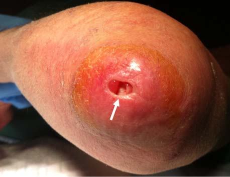
erythema, an elbow wound (figure 1), and generalised
tense oedema and crepitus, the last also being evident on
the ipsilateral supraclavicular region. Plain radiographs
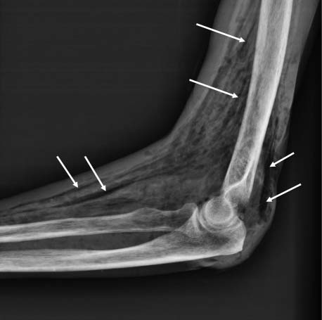
of the left upper limb and chest identified abundant sub-
cutaneous gas (figure 2 and 3), a very specific finding
of necrotising soft-tissue infection (NSTI). The patient
was immediately started on intravenous clindamycin
and vancomycin and then urgent, extensive surgical
Plain radiographs revealing the presence of
subcutaneous widespread gas in the left upper extremity (white
arrows). ‘Dissection’ of muscular and other tissular structures by
debridement of the necrotic tissue was performed. She
afterwards received hyperbaric oxygen as an adjunct to
operative procedure and antibiotics. Culture of several
samples obtained from the necrotic tissue grew no micro-
organisms. The postoperative course was uneventful and
she was discharged on hospital day 11.
NSTI is infrequent but still remains a highly lethal dis-
order.1 Some patients seem to be more prone to develop
this condition, as those with diabetes mellitus, immuno-
suppression, obesity and intravenous drug use.1 2 Other
reported risk factors are age greater than 50 years, periph-
eral vascular disease and chronic alcoholism.2 3 Although
NSTIs are more commonly polymicrobial,1–3 the aetiology
may remain unknown in some patients.1 Early and aggres-
sive surgical debridement combined with empiric broad-
spectrum antimicrobial therapy and physiological support
Left elbow appearance on admission showing a
are of paramount importance to increase the survival
non-exudative, ulcerative lesion (arrow) with surrounded oedema
and erythema (suspected portal of entry for the infection). The
visible portion of the left upper extremity is swollen and
BMJ Case Reports 2012; doi:10.1136/bcr-2012-007025
1. Anaya DA, Dellinger EP. Necrotizing soft-tissue infection: diagnosis and
management. Clin Infect Dis 2007;44:705–10.
2. Ustin JS, Malangoni MA. Necrotizing soft-tissue infections. Crit Care Med
3. Headley AJ. Necrotizing soft tissue infections: a primary care review.
Plain radiographs revealing the presence of
subcutaneous widespread gas in the both ipsilateral hemithorax
(thin black arrows) and supraclavicular area (thick black arrows).
Dissection’ of muscular and other tissular structures by the gas
This pdf has been created automatically from the final edited text and images.
Copyright 2012 BMJ Publishing Group. All rights reserved. For permission to reuse any of this content visit
http://group.bmj.com/group/rights-licensing/permissions.
BMJ Case Report Fellows may re-use this article for personal use and teaching without any further permission.
Please cite this article as follows (you will need to access the article online to obtain the date of publication).
Carrascosa MF, Santamaría MP, Caviedes J-RS, Gutiérrez P G. Necrotising soft-tissue infection. BMJ Case Reports 2012;
Become a Fellow of BMJ Case Reports today and you can:
Enjoy fast sympathetic peer review and rapid publication of accepted articles
▸ Re-use any of the published material for personal use and teaching without further permission
For information on Institutional Fellowships contact consortiasales@bmjgroup.com
Visit casereports.bmj.com for more articles like this and to become a Fellow
BMJ Case Reports 2012; doi:10.1136/bcr-2012-007025
CURRICULUM VITAE Henry (Hank) J. Pieniaszek, Jr., Ph.D., FCP PERSONAL INFORMATION Business Mailing Address: EDUCATION 1978 - 1982 Major: Pharmacy (Pharmaceutical Sciences) Dissertation Topic: Pharmacokinetic Studies with Digitalis Glycosides and Propranolol Dissertation Committee: Department of Pharmaceutical Sciences 1. Donald G. Perrier, Ph.D., co-major advisor 2. M
Anal. Chem. 2004, 76, 4756-4764 Trace Determination of Macrolide and Sulfonamide Antimicrobials, a Human Sulfonamide Metabolite, and Trimethoprim in Wastewater Using Liquid Chromatography Coupled to Electrospray Tandem Mass Spectrometry 1 bel,* Christa S. McArdell, Marc J.-F. Suter, and Walter Giger Swiss Federal Institute for Environmental Science and Technology (EAWAG), CH-8600 Du¨bend
 Miguel F Carrascosa,1 Mariano Pérez Santamaría,2 José-Ramón Salcines Caviedes,1
Department of Internal Medicine, Hospital of Laredo, Laredo, Cantabria, Spain
Service of Orthopedic and Traumatologic Surgery, Hospital of Laredo, Laredo, Cantabria, Spain
Service of Radiology, Hospital of Laredo, Laredo, Cantabria, Spain
Correspondence to Dr Miguel F Carrascosa, miguel.carrascosa@scsalud.es
A 55-year-old woman presented with a 3-day history of
progressively worsening pain, swelling and ‘unpleasant
crackling feeling’ on her left upper limb. These complaints
had begun after she noticed a small reddish lesion on her
left elbow. The patient had received a diagnosis of sys-
temic lupus erythematosus 15 years before coming to us
and was taking methylprednisolone and acenocoumarol,
the last for previous deep vein thrombosis associated with
protein S deficiency. There was history of neither acute or
chronic trauma nor diabetes (in the patient or in her
family). She was allergic to penicillin. On admission,
blood pressure and temperature were normal but heart
rate was 99 beats/min. Her left upper extremity showed
erythema, an elbow wound (figure 1), and generalised
tense oedema and crepitus, the last also being evident on
the ipsilateral supraclavicular region. Plain radiographs
of the left upper limb and chest identified abundant sub-
cutaneous gas (figure 2 and 3), a very specific finding
of necrotising soft-tissue infection (NSTI). The patient
was immediately started on intravenous clindamycin
and vancomycin and then urgent, extensive surgical
Plain radiographs revealing the presence of
subcutaneous widespread gas in the left upper extremity (white
arrows). ‘Dissection’ of muscular and other tissular structures by
debridement of the necrotic tissue was performed. She
afterwards received hyperbaric oxygen as an adjunct to
operative procedure and antibiotics. Culture of several
samples obtained from the necrotic tissue grew no micro-
organisms. The postoperative course was uneventful and
she was discharged on hospital day 11.
Miguel F Carrascosa,1 Mariano Pérez Santamaría,2 José-Ramón Salcines Caviedes,1
Department of Internal Medicine, Hospital of Laredo, Laredo, Cantabria, Spain
Service of Orthopedic and Traumatologic Surgery, Hospital of Laredo, Laredo, Cantabria, Spain
Service of Radiology, Hospital of Laredo, Laredo, Cantabria, Spain
Correspondence to Dr Miguel F Carrascosa, miguel.carrascosa@scsalud.es
A 55-year-old woman presented with a 3-day history of
progressively worsening pain, swelling and ‘unpleasant
crackling feeling’ on her left upper limb. These complaints
had begun after she noticed a small reddish lesion on her
left elbow. The patient had received a diagnosis of sys-
temic lupus erythematosus 15 years before coming to us
and was taking methylprednisolone and acenocoumarol,
the last for previous deep vein thrombosis associated with
protein S deficiency. There was history of neither acute or
chronic trauma nor diabetes (in the patient or in her
family). She was allergic to penicillin. On admission,
blood pressure and temperature were normal but heart
rate was 99 beats/min. Her left upper extremity showed
erythema, an elbow wound (figure 1), and generalised
tense oedema and crepitus, the last also being evident on
the ipsilateral supraclavicular region. Plain radiographs
of the left upper limb and chest identified abundant sub-
cutaneous gas (figure 2 and 3), a very specific finding
of necrotising soft-tissue infection (NSTI). The patient
was immediately started on intravenous clindamycin
and vancomycin and then urgent, extensive surgical
Plain radiographs revealing the presence of
subcutaneous widespread gas in the left upper extremity (white
arrows). ‘Dissection’ of muscular and other tissular structures by
debridement of the necrotic tissue was performed. She
afterwards received hyperbaric oxygen as an adjunct to
operative procedure and antibiotics. Culture of several
samples obtained from the necrotic tissue grew no micro-
organisms. The postoperative course was uneventful and
she was discharged on hospital day 11.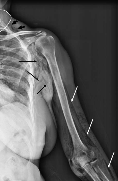 1. Anaya DA, Dellinger EP. Necrotizing soft-tissue infection: diagnosis and
management. Clin Infect Dis 2007;44:705–10.
1. Anaya DA, Dellinger EP. Necrotizing soft-tissue infection: diagnosis and
management. Clin Infect Dis 2007;44:705–10.