Highly efficient transformation of the diatom phaeodactylum tricornutum by multi-pulse electroporation

Biosci. Biotechnol. Biochem., 77 (4), 120936-1–3, 2013 Highly Efficient Transformation of the Diatom Phaeodactylum tricornutumby Multi-Pulse Electroporation Mado MIYAHARA,1 Masaki AOI,1 Natsuko INOUE-KASHINO,2Yasuhiro K 1Graduate School of Biostudies, Kyoto University, Kyoto 606-8502, Japan2Graduate School and Faculty of Science, University of Hyogo, 3-2-1 Kohto, Ako-gun, Hyogo 678-1297, Japan Received December 6, 2012; Accepted January 10, 2013; Online Publication, April 7, 2013 A highly efficient nuclear transformation method was cultured in Daigo’s IMK culture medium (Nihon established for the pennate diatom Phaeodactylum Pharmaceutical, Osaka, Japan) supplemented with sea tricornutum using an electroporation system that drives salts (Sigma, St. Louis, MO) and 0.2 mM Na2SiO3.
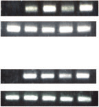
multi-sequence pulses to introduce foreign DNAs into Cultures were grown at 20 C in an artificial climate the cells. By optimizing pulse conditions, the diatom incubator. Cool-white fluorescent tubes provided an cells can be transformed without removing rigid silica- irradiance of 30 mmol photons mÀ2 sÀ1 under continuous based cell walls, and high transformation efficiency (about 4,500 per 108 cells) is achieved.
To generate transgenic P. tricornutum, plasmid pPha- T1, which has a bleomycin-resistant gene cassette with the fcpB promoter,7) was used. The sGFP gene (sgfp) and the GUS (uidA) gene were amplified by PCR usingprimer pairs, 50-GCCGTTTCGAGAATTCATGGTGA- Diatoms are important primary producers View ecosystem that have crucial roles in the global carbon cycle.1) The complex evolutional history of diatoms as 50-AGAATTCTCCCCGGGTGGTCAG-30 and 50-AAAA- secondary endosymbionts is providing new insight into GCTTTGGTGCGCCAGGAGA-30 for uidA. Plasmids host-endosymbiont relationships.2) In addition, their CaMV35S-sGFP(S65T)-nos15) and pBI221 were used as unique abilities to form silica-based cell walls and to templates for PCR. The amplified sgfp and uidA cDNA produce valuable lipids have attracted attention to them HindIII site located in the multi-clonig site of pPha-T1, Both for basic biological research and for commercial which is located downstream of the fcpA promoter.
exploitation of diatoms, an efficient genetic transforma- tion system is very important. Transformation of ditoms (NEPAGENE, Chiba, Japan). Diatom cells at exponen- has been reported for several diatom species, including tial growth phase (OD700 ¼ 0.2–0.4, 5:9 Â 106 cells) pennate diatoms Phaeodactylum tricornutum,5–8) Cylin- were collected by centrifugation at 700 Â g for 4 min, drotheca fusiformis,9,10) and Navicula saprophila, and washed, and resuspended with 0.77 M mannitol. A centric diatoms Cyclothella cryptica,11) Thalassiosira suspension aliquot of 0.15 mL was mixed with 1–10 mg pseudonana,12) and Chaetoceros sp.13) In general, of plasmid that had been linearized by NdeI, and then delivery of foreign DNAs into diatom cells has been transferred to an electroporation cuvette with 0.2 cm achieved by microprojectile bombardment, but this is gap. After electroporation, the cells were transferred into not suited to high-throughput transformation of diatom 4 mL of IMK medium and then incubated to allow cells due to the time required for sample preparation, recovery in nonselective medium at 20 C for 16–20 h costly consumables, and limited transformation efficiency.
under a photon flux density of 30 mmol photons mÀ2 sÀ1.
There is only one report that describes the genetic Considering the slower growth rate of P. tricornutum transformation of P. tricornutum by electroporation,14) cells,16) most of the cells did not be divide during this and this is still at a rudimental stage of development.
incubation period. Cells were collected by centrifugation In this study, we applied a multi-pulse electroporation at 700 Â g over 4 min and resuspended in 0.2 mL of system to achieve nuclear transformation of P. tricor- IMK medium. The transformed cells were selected on nutum. We observed that increases in the number of IMK agar plates containing 1% agar and 100 mg/mL of pulses significantly improved transformation efficiency.
zeocinÒ (Life Technologies, Carlsbad, CA).
To examine the integration of the reporter genes in 108 cells, which is higher than any previously reported the diatom genome, genomic DNA was isolated from transformed cells that were subcultured 2–3 times on P. tricornutum Bohlin (UTEX 642) was derived from selective medium. The protocol described by Kim the University of Texas Culture Collection. It was et al.17) was used to isolate genomic DNA. Primer pairs y To whom correspondence should be addressed. Fax: +81-75-753-6398; E-mail: ifuku@kais.kyoto-u.ac.jpAbbreviations: GFP, green fluorescent protein; GUS, -glucuronidase Illustration of Pulse Regime of Multi-Pulse Electroporation.
Multiple high-voltage poring pulses (P.P.) facilitate the formation of temporary pores, and subsequent multiple low-voltage trans- ferring pulses (T.P.) facilitate the introduction of DNA into the cells.
50-CCGCTTTACTTGTACAGCTCG-30 and 50-CCAT-CTTCTTCAAGGACG-30 for sgfp, 50-GCCACTGGC- Optimization of Pulse Conditions for Electroporation.
(A) Dependence of transformation efficiency on pulse numbers.
Square electric poring pulses were applied at 300 V (pulse duration, 5 ms; 1–9 pulses; interval 50 ms; 10% decay rate), followed by transferring pulses at 8 V (pulse duration, 50 ms; 5 pulses; interval used in genomic PCR. The GFP fluorescence of the 50 ms; 40% decay rate). (B) Dependence of transformation transformed diatom cells was analyzed under a BZ-9000 efficiency on DNA amounts. Square electric poring pulses were fluorescent microscope (KEYENCE, Osaka, Japan). The applied at 300 V (pulse duration, 5 ms; 8 pulses; interval 50 ms; 10% field of cells was excited at 490 nm, and fluorescence decay rate), followed by transferring pulses at 8 V (pulse duration, emission was detected at 510 nm. GUS staining was 50 ms; 5 pulses; interval 50 ms; 40% decay rate).
Figure 1 illustrates a muti-pulse sequence generated examined the buffer for electroporation, and by the electroporation system (NEPA21). Multiple high- found that complete replacement of IMK medium with voltage pulses of short duration and with 10% voltage 0.77 M mannitol gave good results. Furthermore, in- decay (poring pulses) facilitate the formation of tempo- creases in the amount of DNA up to 3–5 mg per cuvette rary pores. Subsequent multiple low-voltage pulses improved transformation efficiency (Fig. 2B). It is very (transferring pulses) of long duration and with 40% important to use a linearized vector to obtain high voltage decay facilitate the delivery of DNA into the transformation efficiency. Without linearization, an cells. During the transfer pulse regime, the electrode approximately 10-fold reduction in transformation effi- polarities are reversed to make the DNA move in the ciency was observed (data not shown). The optimized opposite direction, as this increases transformation transformation efficiency reached about 4,500 transgenic efficiency. This electroporation system was applied for colonies per 108 cells, 10–100 times higher than reported the efficient transformation of various types of animal for other P. tricornutum transformation systems, using cells (http://www.nepagene.jp/E/Eindex.htm).
microprojectile bombardment and the same pPha-T1 This 2-step multi-pulse electroporation was applied to vector.7,8) An additional advantage of the method diatom transformation. It was found that the critical described here is the shorter period required to obtain parameter was the number of poring pulses. For direct zeocin-resistant colonies. Usually it takes 3–4 weeks to comparison of transformation efficiency, we used plas- observe zeocin-resistant colonies by microprojectile mid vector pPha-T1, widely used in previous studies.
bombardment. In our system, zeocin-resistant colonies The GFP (S65T) and the GUS gene (sgfp and uidA started to appear 10–15 d after electroporation. This respectively) were included in pPha-T1 as reporter might have been due to less damage to diatom cells, genes. The voltage of the poring pulses should be higher than 250 V to achieve zeocin-resistant colonies. The We examined the introduction and expression of the pulse duration can be either of 2.5 ms or 5 ms, whereas a sgfp and uidA genes in transgenic diatom cells. It has duration of 5 ms was preferred for higher transformation been reported that some GFP derivatives do not efficiency (data not shown). Figure 2A shows the accumulate efficiently in diatom cells.7) In general, the dependence of the transformation efficiency on the eGFP gene is used for diatom study because it has a number of poring pulses applied. Transformation effi- number of silent mutations that ensure optimum codon ciency reached a maximum when seven pulses were usage in human cells, and they are coincidentally applied, and further increases in the number of pulses preferred by P. tricornuntum. In this study, we used the sGFP (S65T) gene, widely used in plant research.
Highly Efficient Transformation of a Diatom by Electroporation staining in the cells containing uidA (Fig. 3D), confirm- ing that expression of the reporter genes was maintained stably during the repeated segregation process. Expres- 
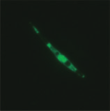
sion level of sgfp in each transgenic line was estimatedfrom the intensity of GFP fluorescence (Fig. 3E). GFP fluorescence levels were different among the transgenic cell lines and this was probably caused by differences in copy number or the position of the reporter gene in thegenome.
In conclusion, the application of the multi-pulse electroporation markedly improved the transformationefficiency of the diatom P. tricornuntum. We found thatthis system was also effective for other algal cells, suchas the green algae Chlamydomonas reinhardtii with cellwalls (Aoi and Ifuku, unpublished data). The methoddescribed here is fast and economical, and shouldcontribute to academic and commercial exploitation ofthe organism.
This work was supported by the Japan Science and Technology Agency, Advanced Low Carbon Technol-ogy Research and Development Program (to K. I. andY. K.).
Falkowski PG, Barber RT, and Smetacek VV, Science, 281,200–207 (1998).
Prihoda J, Tanaka A, de Paula WB, Allen JF, Tirichine L, andBowler C, J. Exp. Bot., 63, 1543–1557 (2012).
Fluorescence (Rel.
TV, Mahapatra DM, Karthick B, and Gordon R, Ind. Eng. Chem. Res., 48, 8769–8788 (2009).
Gordon R, Losic D, Tiffany MA, Nagy SS, and Sterrenburg FA,Trends Biotechnol., 27, 116–127 (2009).
Apt KE, Kroth-Pancic PG, and Grossman AR, Mol. Gen.
Transformant
Falciatore A, Casotti R, Leblanc C, Abrescia C, and Bowler C,Mar. Biotechnol. (NY), 1, 239–251 (1999).
Integration and Expression of the Reporter Genes in Trans- Zaslavskaia LA, Lippmeier JC, Kroth PG, Grossman AR, and genic Phaeodactylum tricornutum Cells.
Apt KE, J. Phycol., 36, 379–386 (2000).
(A) PCR amplification of the sgfp, uidA, and actin genes in Miyagawa A, Okami T, Kira N, Yamaguchi H, Ohnishi K, and transgenic P. tricornutum genomic DNA. Isolated genomic DNAs Adachi M, Phycol. Res., 57, 142–146 (2009).
from the wild type (wt) and four zeocin resistant clones (G1–G4 for Fischer H, Robl I, Sumper M, and Kro¨ger N, J. Phycol., 35, sgfp-transformed cells and U1–U4 for uidA-transformed cells) were used for PCR analysis. Fluorescent (B) and Light (C) photomicro- Poulsen N and Kro¨ger N, FEBS J., 272, 3413–3423 (2005).
graphs of P. tricornutum cells expressing sGFP (S65T). The Du¨nahay TG, Jarvis EE, Zeiler KG, Roessler PG, and Brown presence of the sGFP (S65T) protein was indicated by bright-green LM, Appl. Biochem. Biotechnol., 34–35: 331–339 (1992).
fluorescence. (D) Light photomicrograph of P. tricornutum cells Poulsen N, Chesley PM, and Kro¨ger N, J. Phycol., 42, 1059– expressing GUS. GUS activity was detected as blue staining. Scale bar is 10 mm. (E) Fluorescence levels of transformed P. tricornutum Miyagawa-Yamaguchi A, Okami T, Kira N, Yamaguchi H, cells expressing sGFP. The relative fluorescence levels of the cells in Ohnishi K, and Adachi M, Phycol. Res., 59, 113–119 (2012).
the transgenic lines were estimated by the Image J program.19) Niu YF, Yang ZK, Zhang MH, Zhu CC, Yang WD, Liu JS, and Values are means Æ SD for 5–6 cells.
Li HY, Biotechniques, 0, 1–3 (2012).
Chiu WL, Niwa Y, Zeng W, Hirano T, Kobayashi H, and Sheen Genomic DNA was isolated from transgenic diatom J, Curr. Biol., 6, 325–330 (1996).
cells that had been subcultured 2–3 times on fresh Johnston AM and Raven JA, Mar. Ecol. Prog. Ser., 87, 295–300 selective media after transformation. The presence of the introduced sgfp or uidA gene in the genome was Kim BH, Ramanan R, Cho DH, Choi GG, La HJ, Ahn CY, OhHM, and Kim HS, PLoS ONE, 7, e37770 (2012).
detected by PCR in all the transgenic diatom cells Jefferson RA, Kavanagh TA, and Bevan MW, EMBO J., 6, examined (Fig. 3A). Observation under a fluorescent microscope showed bright GFP fluorescence in the cells Schneider CA, Rasband WS, and Eliceiri KW, Nat. Methods, 9, containing the sgfp gene (Fig. 3B, C) and strong GUS

Source: http://sonidel.com/NEPA21/Highly_Efficient_Transformation_of_the_Diatom_Phaeodactylum-Tricornutum_by_Multi_Pulse_Electroporation.pdf

Zooplancton lagos crÁter de michoacan

Zooplancton epicontinental de los lagos cráter de Michoacán NOMBRE: María Guadalupe Ramos Núñez ESTATUS: Tesista de licenciatura ASESOR: M.C. Rubén Hernández Morales DEPENDENCIA: Laboratorio de Biología Acuática “J. Javier Alvarado Díaz” Facultad INSTITUCIÓN: Universidad Michoacana de San Nicolás de Hidalgo Descripción El zooplancton es una comunidad biol

The havacs cohort description.doc

The HAVACS (HIV Atlanta VA Cohort Study) was initiated in 1982 to clinically care for and study all HIV+ veterans seeking care at the Atlanta VA Medical Center. To date, over 3,000 patients have been entered into this cohort and database. The prospective collection of data was begun in 1982 and includes the full HIV history of patients including any care they received for their HIV disease b

Copyright © 2010-2019 Pdf Physician Treatment