The effect of EDTA, EGTA, EDTAC, and tetracycline-HCl with and without subsequent NaOCl treatment on the microhardness of root canal dentin
Taner Cem Sayin, DDS, PhD,a Ahmet Serper, DDS, PhD,b Zafer C. Cehreli, DDS, PhD,c andHarika G. Otlu, BSc,d Ankara, TurkeyHACETTEPE UNIVERSITY
Objective. The purpose of this study was to evaluate the effect of single and combined use of ethylenediamine tetra acetic acid (EDTA), ethylene glycol bis [b-aminoethylether] N,N,N=,N=-tetraacetic acid (EGTA), EDTA plus Cetavlon (EDTAC), tetracycline-HCl, and NaOCl on the microhardness of root canal dentin. Study design. The crowns of 30 single-rooted human teeth were discarded at the cementoenamel junction and the roots were bisected longitudinally to obtain root halves (N ϭ 60). The specimens were embedded in autopolymerizing acrylic resin, leaving the root canal dentin exposed. Dentin surfaces were prepared for microhardness test by grinding and polishing. The reference microhardness values of untreated specimens were recorded using a Vicker’s microhardness tester at the apical, midroot, and cervical levels of the root canal. Thereafter, the specimens treated with single (test solution only) or combined (test solution, followed by 2.5% NaOCl) versions of the irrigants for 5 minutes. Posttreatment microhardness values were obtained as with initial ones. Statistical comparisons between the test groups and among single and combined treatments were carried out using 2-way ANOVA with repeated measures (P ϭ .05). Comparisons within each group with respect to application regions were made with Friedman’s nonparametric 2-way analysis of variance at the same level of significance. Results. All treatment regimens except distilled water significantly decreased the microhardness of the root canal dentin (P Ͻ . 05). The single and combined use of EDTA decreased the microhardness of the root canal dentin significantly more than all other treatment regimens (P Ͻ .05). Compared with their single-treatment versions, all combined treatment regimens decreased the mean microhardness values significantly (P Ͻ .05). A comparison of single and combined treatment regimens revealed significant decreases only for EDTA and EDTA ϩ NaOCl in the coronal region and for EDTAC and EDTAC ϩ NaOCl in the apical and middle regions of the root canal (P Ͻ .05). Conclusions. The use of EDTA alone or prior to NaOCl resulted in the maximum decrease in dentin microhardness. The softening effect of subsequent NaOCl treatment was both material and region dependent. However, for combined treatment regimens, subsequent use of NaOCl levels the statistical differences between the regional microhardness values obtained after treatment with EGTA, EDTAC, and tetracycline-HCl. (Oral Surg Oral Med Oral Pathol Oral Radiol Endod 2007;104:418-24)
The success of root canal treatment depends on the
ment. However, accumulating evidence suggests the
root canal system being thoroughly cleansed and
importance of removing the smear layer because it
disinfected, followed by obturation of this space.
can result in a more thorough disinfection of the root
Since the first description of the smear layer in
canal system and the dentinal tubules, which would
ensure a better adaptation between the obturation
there is an ongoing debate regarding the influence of
this layer on the success rate of endodontic treat-
created during root canal instrumentation is com-posed of dentin structure and some nonspecific inor-ganic The organic components may
Received from the Faculty of Dentistry, Hacettepe University.
consist of reacted coagulated proteins, necrotic or
aFormerly, Research Assistant, Department of Endodontics. Cur-
viable pulp tissue, odontoblastic processes, and mi-
rently, Assistant Professor, Nova Southeastern University, College of
Dental Medicine, Fort Lauderdale, FL, USA. b
Different solutions have been used to remove the
Professor, Department of Endodontics.
cAssociate Professor, Department of Pediatric Dentistry.
smear layer. Sodium hypochlorite (NaOCl) in a 1%
dResearch Assistant, Department of Biostatistics.
to 5.25% concentration is an irrigant solution widely
Received for publication Oct 12, 2006; returned for revision Mar 7,
used in root canal treatment because of its bacteri-
2007; accepted for publication Mar 19, 2007.
cidal properties and ability to dissolve organic tis-
However, it has been shown to be ineffective
2007 Mosby, Inc. All rights reserved. doi:10.1016/j.tripleo.2007.03.021
in removing the entire smear layer when used
418 419
Thus, the use of chelating agents and acids
MATERIAL AND METHODS
have been suggested to remove the smear layer from
Thirty periodontally involved, human maxillary
the root canal, because the components of this
incisor and mandibular premolar teeth were extracted
loosely bound structure are very small particles with
and stored in distilled water at 4°C for a maximum of
a large surface-mass ratio that makes them very
2 months. Before experiments, soft tissues covering
the root surfaces were removed with gauze and a fine
lating agents are based on different concentrations of
brush. The crowns were removed at the cementoe-
namel junction by using a high-speed bur under
water cooling. Thereafter, the roots were bisected
addition of a quaternary ammonium bromide (Cetavlon)
longitudinally in the buccolingual direction to obtain
increased the action of EDTA by reducing its surface
root halves (N ϭ 60), after which the pulp tissue was
tension, because EDTA solutions act only through direct
removed with a toothbrush. The root halves were
contact with the This combination, known as
embedded in autopolymerizing acrylic resin, leaving
EDTA plus Cetavlon (EDTAC), was shown to be very
the dentin surface exposed. Then, the specimens
effective in smear layer removal and increasing the diam-
were ground flat on a circular grinding machine with
eter of the opened dentinal Recently, Çalt and
ascending grades of SiC abrasive papers (500, 800,
reported that ethylene glycol-bis [b-aminoethyl-
1000, and 1200 grit) under constant water irrigation,
ether]-N,N,N=, N=-tetraacetic acid (EGTA) was also ef-
and further polished with fine alumina suspension
fective in removing the smear layer, without inducing
dentinal erosion commonly caused by EDTA. Tetracy-
The following irrigation solutions were tested in the
cline-hydrochloride (HCl) has also been proposed as a
present study: 2.5% NaOCl, 17% EDTA (ethylenedi-
root canal irrigant. In addition to its antimicrobial effect,
aminetetraacetic acid), 15% EDTAC (EDTA ϩ 0.1%
tetracycline-HCl acts as a calcium chelating agent due to
cationic surfactant, Cetavlon [cetyltrimethylammonium
its low One percent tetracycline HCl has been
bromide]), 17% EGTA (ethylene glycol bis[2-amin-
shown to be as effective as 50% citric acid in the removal
oethylether]-N,N,N=N=-tetraacetic acid), and 1% tetra-
of smear layer, while causing less demineralization in
cycline hydrochloride. All chemicals except NaOCl
were obtained from Sigma Chemical Co. (St. Louis,
It has been reported that some chemicals used for
MO). The test solutions were freshly prepared in lab-
endodontic irrigation are capable of causing alterations in
oratory conditions. The pH of EDTA, EDTAC, EGTA,
the chemical composition of Any change in
and tetracycline-HCl solutions was adjusted to 7.5 by
the Ca/P ratio may alter the original proportion of organic
and inorganic components, which in turn change the mi-
Prior to application of test solutions, the Vicker’s
crohardness, permeability, and solubility characteristics of
hardness values of the specimens were measured on a
Indeed, studies have shown that different con-
Zwick-type 3212002 microhardness tester (Zwick
centrations of EDTA, EDTAC, and EGTA are capable of
GMBH, Ulm, Germany) and recorded. Accordingly, 3
decreasing the microhardness of root canal dentin,
separate indentations, each using 200 gram load and 20
and that this effect can increase by extended application
second dwell time were made along the central axis of
Changes in the mineral content of superficial
the root canal at the apical, midroot, and cervical levels.
dentin may also adversely affect the sealing ability and
The samples were then randomly distributed into the
adhesion of dental materials such as resin-based cements
following treatment groups (n ϭ 10/group): group 1,
2.5% NaOCl; group 2, 17% EDTA; group 3, 17%
For effective removal of both organic and inorganic
EGTA; group 4, 15% EDTAC; group 5, 1% tetracy-
components of the smear layer, it is generally recom-
cline-HCl; and group 6, distilled water (negative con-
mended to use endodontic chelator solutions followed
by Although NaOCl is not a chelating agent,
The specimens were immersed for 5 minutes in a
it can significantly decrease the Ca/P ratio of superficial
magnetic stirrer bath that contained 10 mL of each
test solution. Following treatment with the chelating
on the concentration of the solution. To date, the effect
agents (groups 2 to 5), the same specimens were
of NaOCl on dentin microhardness following initial
treated with NaOCl (combined treatment). Thus,
irrigation with chelating solutions has not been inves-
each specimen served as its own control. In groups 2
tigated. Consequently, the aim of this study was to
to 5, the specimens received a final flush of 10-mL
evaluate the effect of single or combined use of NaOCl,
distilled water immediately after treatment, to avoid
EDTA, EGTA, EDTAC, and tetracycline-HCl on the
the prolonged effect of chelating solutions. The same
microhardness of human root canal dentin.
procedure was carried out after treatment with
420 Table I. Mean of changes (apical ϩ middle ϩ coronal) in the microhardness values of the root canal dentin following treatment with the test solutions Min, minimum; Max, maximum; EDTA, ethylenediamine tetra-aceticacid; EGTA, ethylene glycol-bis [b-aminoethylether]-N,N,N=, N=-tetraacetic acid; EDTAC, EDTA plus Cetavlon.
NaOCl. Posttreatment indentations were made on
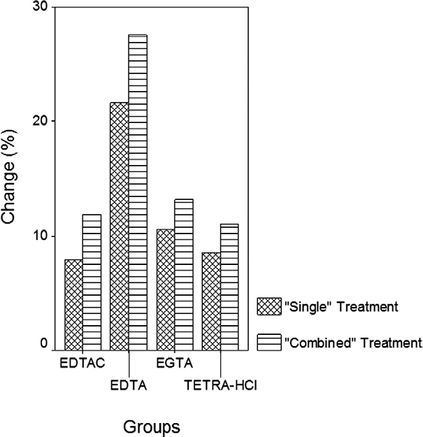
Fig 1. Changes in the microhardness values (percentage)
each specimen adjacent to the initial specimens in the
with respect to single and combined treatment regimens.
same manner, and the microhardness values were
recorded. For each specimen, the change (percent-age) in microhardness values was calculated as fol-lows:
Table II. Changes in microhardness values with re-
spect to apical root canal dentin following treatment
where M ϭ initial microhardness and M ϭ posttreat-
Statistical comparisons between the test groups and
among single and combined treatments were carried out
using 2-way ANOVA with repeated measures (P ϭ
.05). Comparisons within each group with respect to
application regions were made with Friedman’s non-
parametric 2-way analysis of variance at the same level
Min, minimum; Max, maximum; EDTA, ethylenediamine tetra-acetic
acid; EGTA, ethylene glycol-bis [b-aminoethylether]-N,N,N=, N=-
Posttreatment changes in the microhardness values
tetraacetic acid; EDTAC, EDTA plus Cetavlon.
(percentage) of the entire root canal dentin (mean ofapical, middle, and coronal regions) are presented in Changes in the microhardness values (per-centage) with respect to single and combined treat-
3, 4, and 5; P Ͻ .05). However, there was no signif-
icant difference between the microhardness values of
microhardness values with respect to the apical, mid-
EGTA, EDTAC, tetracycline-HCl, and NaOCl (P Ͼ
.05). For single-solution treatments, a statistical
and respectively. All treatment regimens except
ranking for the change in microhardness was ob-
distilled water significantly decreased the microhard-
ness of the root canal dentin (P Ͻ .05). Ethylenedi-
EDTA Ͼ EGTA ϭ EDTAC ϭ tetracycline-HCl
amine tetra-acetic acid decreased the overall micro-hardness of the root canal dentin significantly more
A comparison of combined treatment regimens
than the other single-solution treatments (groups 1,
showed that EDTA ϩ NaOCl induced significantly
421 Table III. Changes in microhardness values with re-
NaOCl and IV; P Ͻ .05). For all single-
spect to middle root canal dentin following treatment
solution treatments, a statistical ranking for the change
in regional microhardness values was obtained as fol-
Min, minimum; Max, maximum; EDTA, ethylenediamine tetra-acetic
In all regions, combined use of EDTA and NaOCl
acid; EGTA, ethylene glycol-bis [b-aminoethylether]-N,N,N=, N=-
decreased the microhardness of the root canal dentin
tetraacetic acid; EDTAC, EDTA plus Cetavlon.
significantly more than EGTA ϩ NaOCl, EDTAC ϩNaOCl, and tetracycline ϩ NaOCl (P Ͻ .05). A com-parison of single and combined treatment regimens
Table IV. Changes in microhardness values with re-
revealed significant decreases only for EDTA and
spect to coronal root canal dentin following treatment
EDTA ϩ NaOCl in the coronal region and for EDTAC
and EDTAC ϩ NaOCl in the apical and middle regions
of the root canal (P Ͻ .05). In all regions, the same
statistical ranking was obtained for changes in micro-
hardness values achieved with combined treatment reg-
ϭ EDTAC ϩ NaOCl ϭ tetracycline-HCl ϩ NaOCl
DISCUSSION
Current concepts of chemomechanical preparation im-
ply that chemicals should be applied on instrumented root
Min, minimum; Max, maximum; EDTA, ethylenediamine tetra-acetic
canal surfaces in order to remove the smear Such
acid; EGTA, ethylene glycol-bis [b-aminoethylether]-N,N,N=, N=-
procedures may induce considerable changes in the
tetraacetic acid; EDTAC, EDTA plus Cetavlon.
surface morphology of dentin, which may also exertchanges in its mechanical and physical proper-Moreover, alteration of the inorganic
more reduction in microhardness than EGTA ϩ
phase of dentin surfaces by acidic pretreatments mod-
NaOCl, EDTAC ϩ NaOCl, and tetracycline ϩ NaOCl
ifies their surface properties, and undoubtedly, their
(P Ͻ .05). When compared with their single-treatment
versions, all combined treatment regimens decreased
correlation between hardness and the mineral content of
the mean microhardness values significantly (P Ͻ .05).
the tooth. The determination of microhardness can thus
For changes in microhardness values achieved with
provide valuable evidence of mineral loss (or gain) in
combined treatment regimens, the following statistical
dental hard with special regard to the effects
In the present study, all specimens were subjected to
a 5-minute contact with the test solutions. Currently,
there is a lack of consensus on the duration a decalci-
EDTAC ϩ NaOCI ϭ tetracycline Ϫ HCI ϩ NaOCI
fying agent must be in contact with the root canal to
With respect to the region being compared (apical,
middle, or coronal), treatment with EDTA resulted in a
herein, De-Deus et limited the contact time of 3
significantly higher decrease in dentin microhardness
chelator solutions (EDTA, EDTAC, and citric acid) to
compared with EGTA, EDTAC, tetracycline-HCl, and
5 minutes, stating that this duration is more realistic in
422
terms of clinical Other researchers have sug-
effective in reducing the surface tension at the apical
gested extending the application time to 10 to 15 min-
region than in the middle and coronal It could
be expected that the removal of the inorganic content of
reported that EDTA can remove the smear layer in 1
dentin would reduce more its microhardness than re-
In addition to contact time, the concentration
move the organic portion. Unlike what is commonly
of the irrigation solution needs to be considered as
accepted, the treatment of dentin with NaOCl may not
another determinant in the posttreatment microhardness
only remove the organic matrix but also some of the
values of dentin. On the basis of the results obtained,
inorganic content that ultimately renders dentin much
EDTA decreased the microhardness of dentin by
weaker than The precise mechanism of this
17.33% to 29.48%, and this effect was significantly
phenomenon is unknown, leaving room for speculation.
greater than that achieved with both the test and control
With special regard to the combined treatment regi-
solutions. Although EDTA and EDTAC had similar
mens tested, subsequent application of NaOCl may
concentrations (17% vs. 15%), the efficacy of EDTAC
facilitate further exposure of the inorganic material on
was significantly lower than that of EDTA. This finding
decalcified dentin substrate through removal of the
corroborates previous work,showing that reduc-
ing its surface tension does not improve the effective-
izing effect that would eventually decrease the dentin
ness of EDTA. According to De-Deus et the lesser
microhardness. Nevertheless, this effect can be material
efficiency of EDTAC to remove calcium ions from
and/or region dependent. For instance, compared with
dentin could be responsible for this finding. Although
their respective single-treatment versions, significant
this explanation could be reasonably extended to the
microhardness reductions in the combined treatment
findings obtained with EGTA and tetracycline-HCl,
groups were observed only when NaOCl was used after
there is currently no published study to support this
EDTA in the coronal third and after EDTAC in the
assumption, especially when all solutions are adjusted
apical and middle thirds of the root canal.
to the same concentration and/or pH.
Microhardness tests have been traditionally em-
The relative softening effect on dentinal walls ex-
ployed to evaluate materials, presenting a certain ho-
erted by chemical irrigants could be of clinical benefit
Biological materials such as dentin are far
since it permits rapid preparation and facilitates nego-
less homogenous, with dentin tubule density increasing
tiation of small tight but these alterations
from cervical to apical resulting in an inverse
also affect the sealing ability and adhesion of sealers to
correlation between dentin microhardness and tubule
This may lead to deviations in the results
solution removes the entire smear within the same
because of differences in adjacent regions of the dentin
period of time, lower concentrations of EDTA should
This is clearly confirmed in the present study
be preferred to reduce its adverse (softening) effect on
by the differences in the statistical ranking of single-
root dentin. In this regard, the tested concentrations of
solution treatments with EGTA, EDTAC, NaOCl, and
EGTA and EDTAC can be considered less detrimental
tetracycline-HCl at the apical, middle, and coronal re-
to dentin. However, the efficacy of lower concentra-
gions of root canal dentin. However, following subse-
tions of EGTA and EDTAC merits further evaluation,
quent treatment with NaOCl, the statistical ranking for
because these 2 solutions also significantly decreased
all three regions was the same. This indicates that
regional differences in microhardness are leveled in the
ing solutions also needs to be considered as another
combined treatment groups in a similar pattern ob-
important factor. However, since the pH of all test
served in the general mean ranking of all 3 regions
solutions were adjusted to 7.5, comparisons cannot be
suggests that the combination of tetracycline-HCl and
Results obtained within the experimental conditions
NaOCl appears to yield the least softening (adverse)
of the present study indicate that the single use of
NaOCl significantly reduces the microhardness of rootcanal dentin compared with control. Further, despite the
CONCLUSION
lack of significant differences, comparison of numerical
On the basis of the results obtained and experimental
data has shown that the use of NaOCl alone can also
conditions of the present study, the use of EDTA alone
induce more reduction in microhardness in comparison
or prior to NaOCl resulted in the maximum decrease in
with EDTAC and tetracycline-HCl in the middle and
dentin microhardness. The softening effect of subse-
coronal root canal dentin. NaOCl was not as effective
quent NaOCl treatment was both material and region
in the apical region as it was in the coronal and middle
dependent. However, for combined treatment regimens,
thirds, probably because it has been shown to be less
subsequent use of NaOCl levels the differences be-
423
tween the microhardness values obtained after treat-
of citric acid solutions on the calcium and phosphorus contents of
ment with EGTA, EDTAC, and tetracycline-HCl.
human root dentin. J Endod 1994;20:551-4.
23. Rotstein I, Dankner E, Goldman A, Heling I, Stabholz A, Zalkind
M. Histochemical analysis of dental hard tissues following
REFERENCES
1. McComb D, Smith DC. A preliminary scanning electron micro-
24. Ari H, Erdemir A. Effects of endodontic irrigation solutions on
scopic study of root canals after endodontic procedures. J Endod
mineral content of root canal dentin using ICP-AES technique.
2. Oksan T, Aktener BO, Sen BH, Tezel H. The penetration of root
25. Cruz-Filho AM, Sousa-Neto MD, Saquy PC, Pecora JD. Evalu-
canal sealers into dentinal tubules. A scanning electron micros-
ation of the effect of EDTAC, CDTA, and EGTA on radicular
copy study. Int Endod J 1993;26:301-5.
dentin microhardness. J Endod 2001;27:183-4.
3. Kouvas V, Liolios E, Vassiliadis L, Parissis-Messimeris S, Bout-
26. Perinka L, Sano H, Hosoda H. Dentin thickness, hardness and
sioukis A. Influence of smear layer on depth of penetration of
Ca-concentration vs. bond strength of dentin adhesives. Dent
three endodontic sealers: a SEM study. Endod Dent Traumatol
27. Panighi M, G Sell C. Influence of calcium concentration on the
4. Clark-Holke D, Drake D, Walton R, Rivera E, Guthmiller JM.
dentin wettability by an adhesive. J Biomed Mater Res1992;26:1081-9.
Bacterial penetration through canals of endodontically treated
28. Perdigao J, Eiriksson S, Rosa BT, Lopes M, Gomes G. Effect of
teeth in the presence or absence of the smear layer. J Dent
calcium removal on dentin bond strengths. Quintessence Int
5. Pashley DH. Smear layer: overview of structure and function.
29. Garcia-Godoy F, Loushine RJ, Itthagarun A, Weller RN, Murray
Proc Finn Dent Soc 1992;88(Suppl l):215-24.
PE, Feilzer AJ, et al. Application of biologically-oriented dentin
6. Czonstkowsky M, Wilson EG, Holstein FA. The smear leayer in
bonding principles to the use of endodontic irrigants. Am J Dent
endodontics. Dent Clin North Am 1990;34:13-25.
7. Yamada RS, Armas A, Goldman M, Lin PS. A scanning electron
30. Slutzky-Goldberg I, Maree M, Liberman R, Heling I. Effect of
microscopic comparison of a high volume final flush with several
irrigating solutions: part 3. J Endod 1983;9:137-42.
8. Baumgartner JC, Mader CL. A scanning electron microscopic
31. Ari H, Erdemir A, Belli S. Evaluation of the effect of endodontic
evaluation of four root canal irrigation regimens. J Endod
irrigation solutions on the microhardness and the roughness of
root canal dentin. J Endod 2004;30:792-5.
9. Prati C, Selighini M, Ferrieri P, Mongiorgi R. Scanning electron
32. Grigoratos D, Knowles J, Ng YL, Gulabivala K. Effect of exposing
microscopic evaluation of different endodontic procedures on
dentine to sodium hypochlorite and calcium hydroxide on its flex-
dentin morphology of human teeth. J Endod 1994;20:174-9.
ural strength and elastic modulus. Int Endod J 2001;34:113-9.
10. Berutti E, Marini R. A scanning electron microscopic evaluation
33. Sim TP, Knowles JC, Ng YL, Shelton J, Gulabivala K. Effect of
of the debridement capability of sodium hypochlorite at different
sodium hypochlorite on mechanical properties of dentine and
temperatures. J Endod 1996;22:467-70.
tooth surface strain. Int Endod J 2001;34:120-32.
11. Bertrand MF, Pizzardini P, Muller M, Medioni E, Rocca JP. The
34. Van Meerbeek B, Willems G, Celis JP, Roos JR, Braem M,
removal of the smear layer using the quantec system. A study using
Lambrechts P, et al. Assessment by nano-indentation of the
the scanning electron microscope. Int Endod J 1999;32:217-24.
hardness and elasticity of the resin-dentin bonding area. J Dent
12. Torabinejad M, Handysides R, Khademi AA, Bakland LK. Clinical
implications of the smear layer in endodontics: a review. Oral Surg
35. Panighi M, G Sell C. Effect of the tooth microstructure on the
Oral Med Oral Pathol Oral Radiol Endod 2002;94:658-66.
shear bond strength of a dental composite. J Biomed Mater Res
13. De-Deus G, Paciornik S, Mauricio MH. Evaluation of the effect
of EDTA, EDTAC and citric acid on the microhardness of root
36. Arends J, ten Bosch JJ. Demineralization and remineralization
evaluation techniques. J Dent Res 1992;71:924-8.
14. Dogan H, Calt S. Effects of chelating agents and sodium hypochlo-
37. Calt S, Serper A. Time-dependent effects of EDTA on dentin
rite on mineral content of root dentin. J Endod 2001;27:578-80.
15. Hulsmann M, Heckendorff M, Lennon A. Chelating agents in
38. Scelza MF, Pierro V, Scelza P, Pereira M. Effect of three
root canal treatment: mode of action and indications for their use.
different time periods of irrigation with EDTA-T, EDTA and
citric acid on smear layer removal. Oral Surg Oral Med Oral
16. Hill PK. Endodontics. J Prosthet Dent 1959;9:142.
Pathol Oral Radiol Endod 2004;98:499-503.
17. Goldberg F, Abramovich A. Analysis of the effect of EDTAC on
39. Zehnder M, Schicht O, Sener B, Schmidlin P. Reducing surface
the dentinal walls of the root canal. J Endod 1977;3:101-5.
tension in endodontic chelator solutions has no effect on their
18. Goldberg F, Spielberg C. The effect of EDTAC and the variation
ability to remove calcium from instrumented root canals. J Endod
of its working time analyzed with scanning electron microscopy.
Oral Surg Oral Med Oral Pathol 1982;53:74-7.
40. Saleh AA, Ettman WM. Effect of endodontic irrigation solutions
19. Calt S, Serper A. Smear layer removal by EGTA. J Endod
on microhardness of root canal dentine. J Dent 1999;27:43-6.
41. Cruz-Filho AM, Paula EA, Pecora JD, Sousa-Neto MD. Effect of
20. Wikesjo UM, Baker PJ, Christersson LA, Genco RJ, Lyall RM,
different EGTA concentrations on dentin microhardness. Braz
Hic S, et al. A biochemical approach to periodontal regeneration:
tetracycline treatment conditions dentin surfaces. J Periodontal
42. Pecora JD, Sousa-Neto MD, Guerisoli DMZ, Marchesan MA.
Effect of reduction of the surface tension of different concentra-
21. Haznedaroglu F, Ersev H. Tetracycline HCl solution as a root
tions of sodium hypochlorite solutions on radicular dentine per-
canal irrigant. J Endod 2001;27:738-40.
meability. Braz Endod J 1998;3:38-40.
22. Hennequin M, Pajot J, Avignant D. Effects of different pH values
43. Sakae T, Mishima H, Kozawa Y. Changes in bovine dentin
424
mineral with sodium hypochlorite treatment. J Dent Res
47. Pashley D, Okabe A, Parham P. The relationship between dentin mi-
crohardness and tubule density. Endod Dent Traumatol 1985;1:176-9.
44. Inaba D, Ruben J, Takagi O, Arends J. Effect of sodium hypo-
chlorite treatment on remineralization of human root dentine in
45. Niu W, Yoshioka T, Kobayashi C, Suda H. A scanning electron
microscopic study of dentinal erosion by final irrigation with
EDTA and NaOCl solutions. Int Endod J 2002;35:934-9.
46. Carrigan PJ, Morse DR, Furst ML, Sinai IH. A scanning electron
microscopic evaluation of human dentinal tubules according to
age and location. J Endod 1984;10:359-63.
Los productos de la industria de los pesticidas controlan el desarrollo de insectos, roedores o moluscos. Se emplean en áreas urbanas, suburbanas y rurales; en el hogar y en los jardines. La industria utiliza herbicidas, alguicidas, funguicidas y bactericidas y los ferrocarriles usan herbicidas para mantener las vías libres de vegetación. La necesidad de un nombre genérico que englobe la gra
 420
420